
Page 1193 - Equine Clinical Medicine, Surgery and Reproduction, 2nd Edition
P. 1193
1168 CHAPTER 11
VetBooks.ir 11.74 11.75
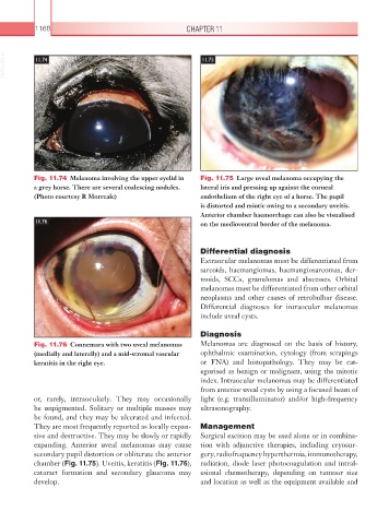
Fig. 11.74 Melanoma involving the upper eyelid in Fig. 11.75 Large uveal melanoma occupying the
a grey horse. There are several coalescing nodules. lateral iris and pressing up against the corneal
(Photo courtesy R Morreale) endothelium of the right eye of a horse. The pupil
is distorted and miotic owing to a secondary uveitis.
Anterior chamber haemorrhage can also be visualised
11.76 on the medioventral border of the melanoma.
Differential diagnosis
Extraocular melanomas must be differentiated from
sarcoids, haemangiomas, haemangiosarcomas, der-
moids, SCCs, granulomas and abscesses. Orbital
melanomas must be differentiated from other orbital
neoplasms and other causes of retrobulbar disease.
Differential diagnoses for intraocular melanomas
include uveal cysts.
Diagnosis
Fig. 11.76 Connemara with two uveal melanomas Melanomas are diagnosed on the basis of history,
(medially and laterally) and a mid-stromal vascular ophthalmic examination, cytology (from scrapings
keratitis in the right eye. or FNA) and histopathology. They may be cat-
egorised as benign or malignant, using the mitotic
index. Intraocular melanomas may be differentiated
from anterior uveal cysts by using a focused beam of
or, rarely, intraocularly. They may occasionally light (e.g. transilluminator) and/or high-frequency
be unpigmented. Solitary or multiple masses may ultrasonography.
be found, and they may be ulcerated and infected.
They are most frequently reported as locally expan- Management
sive and destructive. They may be slowly or rapidly Surgical excision may be used alone or in combina-
expanding. Anterior uveal melanomas may cause tion with adjunctive therapies, including cryosur-
secondary pupil distortion or obliterate the anterior gery, radiofrequency hyperthermia, immunotherapy,
chamber (Fig. 11.75). Uveitis, keratitis (Fig. 11.76), radiation, diode laser photocoagulation and intral-
cataract formation and secondary glaucoma may esional chemotherapy, depending on tumour size
develop. and location as well as the equipment available and