
Page 1288 - Equine Clinical Medicine, Surgery and Reproduction, 2nd Edition
P. 1288
Skin 1263
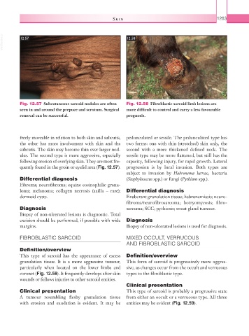
VetBooks.ir 12.57 12.58
Fig. 12.57 Subcutaneous sarcoid nodules are often Fig. 12.58 Fibroblastic sarcoid limb lesions are
seen in and around the prepuce and scrotum. Surgical more difficult to control and carry a less favourable
removal can be successful. prognosis.
freely moveable in relation to both skin and subcutis, pedunculated or sessile. The pedunculated type has
the other has more involvement with skin and the two forms: one with thin (stretched) skin only, the
subcutis. The skin may become thin over larger nod- second with a more thickened defined neck. The
ules. The second type is more aggressive, especially sessile type may be more flattened, but still has the
following erosion of overlying skin. They are most fre- capacity, following injury, for rapid growth. Lateral
quently found in the groin or eyelid area (Fig. 12.57). progression is by local invasion. Both types are
subject to invasion by Habronema larvae, bacteria
Differential diagnosis (Staphylococcus spp.) or fungi (Pythium spp.).
Fibroma; neurofibroma; equine eosinophilic granu-
loma; melanoma; collagen necrosis (axilla – rare); Differential diagnosis
dermoid cysts. Exuberant granulation tissue; habronemiasis; neuro-
fibroma/neurofibrosarcoma; botryomycosis; fibro-
Diagnosis sarcoma; SCC; pythiosis; sweat gland tumour.
Biopsy of non-ulcerated lesions is diagnostic. Total
excision should be performed, if possible with wide Diagnosis
margins. Biopsy of non-ulcerated lesions is used for diagnosis.
FIBROBLASTIC SARCOID MIXED OCCULT, VERRUCOUS
AND FIBROBLASTIC SARCOID
Definition/overview
This type of sarcoid has the appearance of excess Definition/overview
granulation tissue. It is a more aggressive tumour, This form of sarcoid is progressively more aggres-
particularly when located on the lower limbs and sive, as changes occur from the occult and verrucous
coronet (Fig. 12.58). It frequently develops after skin types to the fibroblastic type.
wounds or follows injuries to other sarcoid entities.
Clinical presentation
Clinical presentation This type of sarcoid is probably a progressive state
A tumour resembling fleshy granulation tissue from either an occult or a verrucous type. All three
with erosion and exudation is evident. It may be entities may be evident (Fig. 12.59).