
Page 1302 - Equine Clinical Medicine, Surgery and Reproduction, 2nd Edition
P. 1302
Skin 1277
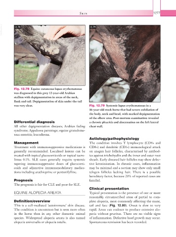
VetBooks.ir 12.78 12.79
Fig. 12.78 Equine cutaneous lupus erythematosus
was diagnosed in this grey 12-year-old Arabian
stallion with depigmentation in areas of the neck,
flank and tail. Depigmentation of skin under the tail
was very clear. Fig. 12.79 Systemic lupus erythematosus in a
16-year-old stock horse that had severe exfoliation of
the body, neck and head, with marked depigmentation
of the elbow area. Post-mortem examination revealed
Differential diagnosis a chronic pleuritis and abscessation on the left lateral
All other depigmentation diseases; Arabian fading chest wall.
syndrome; Appaloosa parentage; equine granuloma-
tous enteritis; leucoderma.
Aetiology/pathophysiology
Management The condition involves T lymphocyte (CD4+ and
Treatment with immunosuppressive medications is CD8+) and dendritic (CD1+) immunological attack
generally recommended. Localised lesions can be on anagen hair follicles, characterised by antibod-
treated with topical glucocorticoids or topical tacro- ies against trichohyalin and the inner and outer root
limus 0.1%. SLE cases generally require systemic sheath. Early diseased hair follicles may show defec-
tapering immunosuppressive doses of glucocorti- tive keratinisation. In chronic cases, inflammation
coids and adjunctive immunomodulatory medica- may be minimal and a section may show only small
tions including azathioprine or pentoxifylline. telogen follicles lacking hair. There is a possible
hereditary factor, because 20% of reported cases are
Prognosis familial.
The prognosis is fair for CLE and poor for SLE.
Clinical presentation
EQUINE ALOPECIA AREATA Typical presentation is the presence of one or more
reasonably circumscribed areas of partial to com-
Definition/overview plete alopecia, most commonly affecting the mane,
This is a cell-mediated ‘autoimmune’ skin disease. tail and face (Fig. 12.80). Onset is slow to very
The condition is uncommon but is seen more often rapid. Areas can coalesce to produce extensive alo-
in the horse than in any other domestic animal pecia without pruritus. There are no visible signs
species. Widespread alopecia areata is also named of inflammation. Defective hoof growth may occur.
alopecia universalis or alopecia totalis. Spontaneous remission has been recorded.