
Page 188 - Equine Clinical Medicine, Surgery and Reproduction, 2nd Edition
P. 188
Musculoskeletal system: 1.4 The forelimb 163
VetBooks.ir Full carpal flexion may be restricted with marked Radiography can rule out any bony involvement
and contrast medium is used to investigate joint or
soft-tissue swelling. If the hygroma is infected, the
swelling may be more pronounced, with serous ooz-
ing and pain present. tendon sheath involvement.
Management
Differential diagnosis Conservative treatment involves rest, local injections
Extensor tendon sheath effusion; carpal joint of steroids, drainage and bandaging. Although in some
effusion/herniation. cases these may resolve the swelling, conservative treat-
ment is usually unsuccessful. Surgical treatment requires
Diagnosis en bloc resection of the tissue, avoiding penetration of
Careful clinical palpation and knowledge of anat- the extensor tendon sheath or joint capsule, followed by
omy are required to differentiate carpal hygroma a sleeve cast or Robert Jones bandage for 7–10 days.
from effusions of the extensor tendon sheaths or
carpal joints. Ultrasonography is useful for exam- Prognosis
ining the hygroma and other nearby structures The prognosis is guarded for complete resolution as
and looking for the possibility of a foreign body. recurrence is common.
ANTEBRACHIUM AND ELBOW
CARPAL CANAL SYNDROME depending on the underlying cause, and flexion of
the carpus usually exacerbates the clinical signs.
Definition/overview
Carpal canal syndrome involves conditions lead- Differential diagnosis
ing to restriction or pain as the carpal sheath passes Carpal joint pathology; extensor tendon sheath
through the carpal region. pathology.
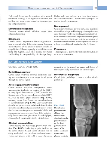
Aetiology/pathophysiology 1.305
Causes include idiopathic tenosynovitis, septic
tenosynovitis, tendinitis or tearing of the SDFT
or deep digital flexor tendon (DDFT)/muscle bel-
lies, desmitis of the accessory ligament of the SDFT
(AL-SDFT), radial physis exostosis (Fig. 1.305),
accessory carpal bone fracture or osteochondroma
of the distal radius (Fig. 1.306). Osteochondromas
describe a separate area of endochondral ossification Fig. 1.305
from the caudal (usually caudomedial) aspect of the Dorsolateral/
distal radius, resulting in irritation of the DDFT and palmaromedial
accompanying tenosynovitis. These differ histologi- oblique view
cally from exostosis or spikes from the radial physis, of the carpus
although both can result in similar clinical signs. showing irregular
protruberances
Clinical presentation on the distal
Horses usually, but not always, present with effu- palmarolateral
sion (Fig. 1.307), thickening or pain localised to aspect of the
the carpal sheath. Carpal sheath effusion can be radius consistent
easily overlooked, particularly on the lateral aspect. with a radial
Horses can present with mild to severe lameness physeal exostosis.