
Page 200 - Equine Clinical Medicine, Surgery and Reproduction, 2nd Edition
P. 200
Musculoskeletal system: 1.4 The forelimb 175
VetBooks.ir OSTEOCHONDROSIS Diagnosis
Diagnosis can be suspected from clinical signs
Definition/overview
in the shoulder may show a range of signs from
Conditions that affect the scapulohumeral joint and signalment. Radiography of horses with OCD
include OCD of the humeral head and glenoid cav- mild flattening of the humeral head and subchon-
ity and OCLLs of the glenoid cavity of the scapula dral bone lucency, through to overt mineralised
(and rarely humerus). flaps. Contrast radiography may further delin-
eate flaps but often radiographic signs underplay
Aetiology/pathophysiology the severity of the condition (Fig. 1.335). Signs
OCD of the shoulder joint is less common than in of secondary OA are common. Ultrasonography
other locations but has a similar aetiology. may be helpful in confirming joint effusion
and osteochondral flaps can also be visual-
Clinical presentation ised, particularly in the caudal part of the joint.
Horses with OCD of the shoulder joint may pres- OCLLs may be of variable size and located on
ent relatively late (yearlings or older) compared the glenoid rim with or without a sclerotic rim
with other sites (Fig. 1.334). Lameness is usually (Fig. 1.336). Nuclear scintigraphy may highlight
moderate-severe with evidence of muscle atrophy and, increased radiopharmaceutical uptake with an
in some cases, joint effusion. Pain on manipulation is OCLL.
present and some horses may present with secondary
contracture of the DIP joint (clubbed or boxy foot) Management
in chronic cases. Horses with OCLLs are generally Arthroscopic surgery is advised to remove dis-
older and can present with lameness of an intermit- secting cartilage flaps and to curette underlying
tent nature and severity. Clinical signs may suggest subchondral bone (Figs. 1.337, 1.338). Severe
shoulder pain, but often localising signs are scant. cases may present with complete lifting off of the
cartilage. Although pinning techniques have been
Differential diagnosis described, euthanasia in severe cases is recom-
Shoulder OA; dysplasia; luxation; synovial sepsis. mended due to the rapid development of secondary
1.334 1.335
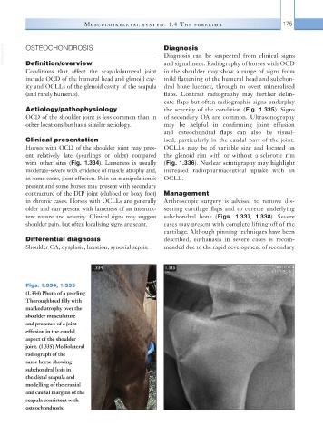
Figs. 1.334, 1.335
(1.334) Photo of a yearling
Thoroughbred filly with
marked atrophy over the
shoulder musculature
and presence of a joint
effusion in the caudal
aspect of the shoulder
joint. (1.335) Mediolateral
radiograph of the
same horse showing
subchondral lysis in
the distal scapula and
modelling of the cranial
and caudal margins of the
scapula consistent with
osteochondrosis.