
Page 253 - Equine Clinical Medicine, Surgery and Reproduction, 2nd Edition
P. 253
228 CHAPTER 1
VetBooks.ir 1.430 usually stabilised by the contralateral mandible or
Unilateral fractures of the interdental space are
maxilla, and therefore are often dealt with conserva-
tively. Bilateral maxillary fractures (Fig. 1.431) may
also involve trauma to the nasal septum, nasal pro-
cess, facial bones and hard palate, but in many cases
may heal successfully without fixation. Bilateral
mandibular fractures involving the interdental space
are often completely unstable and demand fixation,
which can be achieved using a variety of techniques.
Tension-band wires from the incisors to the cheek
teeth, U-shaped frames held by wires around the
teeth, frames using pins through the mandible or,
more recently, using pinless external fixation devices
have all been described as standalone techniques or
in combination. Most methods are performed with
the horse under general anaesthesia, but some tech-
niques (and patients) lend themselves to standing sur-
gery using sedation and perineural local anaesthesia.
Internal fixation using bone plates is uncommon, but
is indicated for those fractures of the caudal man-
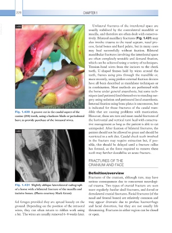
Fig. 1.430 A groove cut in the caudal aspect of the dible that are causing problems with mastication.
canine (204) tooth, using a hacksaw blade or periodontal However, these are rare and most caudal fractures of
burr, to provide purchase of the intraoral wires. the horizontal and vertical rami heal with conserva-
tive management as long as the patient is able to eat
unimpeded. After fixation of bilateral fractures, the
1.431 patient should not be allowed to graze and should be
restricted to a soft diet. Caudal cheek teeth involved
in the fracture may require extraction but, if pos-
sible, this should be delayed until a fracture callus
has formed, as the force required to remove these
teeth may further destabilise an acute fracture.
FRACTURES OF THE
CRANIUM AND FACE
Definition/overview
Fractures of the cranium, although rare, may have
serious consequences due to concurrent neurologi-
Fig. 1.431 Slightly oblique laterolateral radiograph cal trauma. Two types of cranial fracture are seen
of a horse with a bilateral fracture of the maxilla and more regularly: basilar skull fractures, and dorsal or
incisive bones. (Photo courtesy Mark Grant) dorsolateral cranial fractures. Facial fractures (of the
nasal and frontal bones) are relatively common and
fed forages provided they are spread loosely on the may appear dramatic due to profuse haemorrhage
ground. Depending on the position of the intraoral and facial distortion, but they are not usually life
wires, they can often return to ridden work using threatening. Fractures in either region can be closed
a bit. The wires are usually removed 6–8 weeks later. or open.