
Page 276 - Equine Clinical Medicine, Surgery and Reproduction, 2nd Edition
P. 276
Musculoskeletal system: 1.7b The axial skeleton – thoracolumbar region 251
VetBooks.ir 1.7b The axial skeleton – thoracolumbar region
BACK ANATOMY AND FUNCTION is normally complete shortly after birth, but sepa-
rate centres of ossification, such as in the extremi-
The thoracolumbar spine is the central part of the ties of the transverse and dorsal spinous processes, as
horse’s musculoskeletal core, which is made up of well as the epiphyses of the lumbar vertebral bodies,
the neck, back, sacral area and tail. Lameness prob- remain present for many years (Fig. 1.472). Those
lems can alter spinal function through compensa- in the dorsal spinous processes (DSPs) are reported
tion to abnormal movements elsewhere in the body. to close between 9 and 14 years of age but can be
Progressive chronic problems of the thoracolumbar found even later (Fig. 1.473). Lumbar vertebral
region cannot therefore be considered in isolation body physes may take between 5 and 7 years to close.
from the remainder of the core or from the muscu-
loskeletal system as a whole. Soft-tissue function
Each vertebra contacts its neighbours via a single
Axial skeleton fibrocartilage intervertebral disc separating the ver-
The horse has a vertebral formula of 18 thoracic and tebral body and via left and right articular processes,
six lumbar vertebrae. The size and shape of vertebrae which form a facet joint on each side (Figs. 1.474–
changes gradually from cranial to caudal based on 1.476). Several ligaments of varying length link the
their function within the back. Primary ossification bones together. The supraspinous and interspinous
1.472
Fig. 1.472 Lateral radiograph of the
caudal thoracic region of a 3-week-
old foal. The bones are formed
but epiphyses remain open in the
vertebral bodies and the dorsal tips of
the dorsal spinous processes are not
ossified.
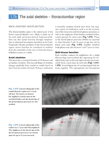
1.473
Fig. 1.473 Lateral radiograph of the
withers in a 7-year-old Thoroughbred.
The epiphyses in the dorsal tips of the
dorsal spinous processes can remain
open for up to 14 years (arrows).