
Page 301 - Equine Clinical Medicine, Surgery and Reproduction, 2nd Edition
P. 301
276 CHAPTER 1
VetBooks.ir Differential diagnoses cutaneously and rectally (Figs. 1.521–1.523). The
Ultrasound images can be obtained both trans-
Hindlimb lameness; lumbosacral or thoracolumbar
pain; stress fractures of the ilial wing adjacent to the
SI joint. dorsal part of the dorsal SI ligament and bony
margins of the sacrum and ilium can be obtained
by imaging transcutaneously, from a dorsoventral
Diagnosis direction, on either side of the sacral spinous pro-
Radiographic evaluation of the SI region is difficult cesses. The ventral ligament can be imaged via a
due to the superimposition of large muscles and transrectal approach, which can also be used to
abdominal viscera. High-quality radiography of the determine the angle and width of the joint space.
SI joints requires general anaesthesia and is conse- The dorsal portion of the dorsal ligament appears
quently seldom performed. to be most commonly affected, showing alterations
in size, loss of normal echogenicity and parallel fibre
1.520 1.521
Fig. 1.521 Transcutaneous ultrasound image
of a lesion in the short dorsal sacroiliac ligament;
transverse view (left) and longitudinal view (right).
(Photo courtesy Diane Isbell
1.522
Sacrum
Ilium
R Joint
Ventral
ligament
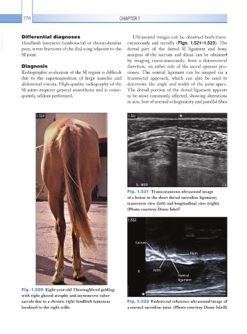
Fig. 1.520 Eight-year-old Thoroughbred gelding
with right gluteal atrophy and asymmetric tuber
sacrale due to a chronic right hindlimb lameness Fig. 1.522 Endorectal reference ultrasound image of
localised to the right stifle. a normal sacroiliac joint. (Photo courtesy Diane Isbell)