
Page 539 - Equine Clinical Medicine, Surgery and Reproduction, 2nd Edition
P. 539
514 CHAPTER 2
VetBooks.ir fibrous nodule attaching the tail of the epididymis albuginea and enter the head of the epididymis. This
results in a mediastinum testis that is less prominent
to the caudal pole of the testicle. It can be relatively
large in newborn colts and, on palpation, may be
sonographically (Fig. 2.122).
mistaken for a testicle within the scrotum. On occa- grossly on cut section and almost indefinable ultra-
sion, examination of a normal stallion may iden-
tify rotation of up to 180° of one or both testicles. EPIDIDYMIDES AND EXCURRENT
Testicle rotation is often transient and a subsequent DUCT SYSTEM
examination may find the testicle in normal orienta-
tion. Rotation must be differentiated from true tes- Each epididymis is a highly convoluted, but
ticular or spermatic cord torsion, in which stallions unbranched, duct approximately 70 metres long and
demonstrate signs of colic and palpation reveals a having a grossly distinct head, body and tail. In the
painful and swollen testicle. stallion, the head of the epididymis is a flattened
The testis is encapsulated by the tunica albuginea, structure that lies dorsomedially along the cranial
a layer of tough collagenous tissue and smooth mus- border of the testis. The body, or corpus, lies along
cle that is fused to the visceral layer of the vaginal the dorsolateral aspect of each testis and continues as
tunic. The tunica albuginea sends supportive tra- the tail, or cauda, which is a large, prominent struc-
beculae into the testicular parenchyma, dividing the ture attached to the caudal pole of the testis. The
testis into lobules. The testicle of the stallion does deferent duct, the excretory duct for sperm, attaches
not contain an axially oriented mediastinum testis as to the tail of the corresponding epididymis, runs
is seen in the bull and other species. In the stallion, along the medial aspect of the testis and ascends via
it is located at the cranial pole of the testis, where the the spermatic cord through the vaginal ring into the
excurrent ducts leaving the testis cross the tunica pelvis. Each deferent duct widens into its correspond-
ing ampullary gland and eventually terminates at the
colliculus seminalis of the pelvic urethra. The collic-
2.122 ulus seminalis is a rounded prominence situated on
+ the dorsomedial wall of the urethra about 5 cm cau-
dal to the urethral opening from the bladder. This is
the site at which the ducts of the accessory sex glands
Central vein empty into the urethra. Whereas the deferent ducts
2.123
Right testis
1
+
Testis
Tail of epididymis
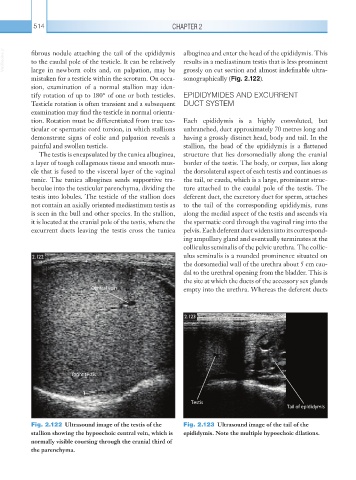
Fig. 2.122 Ultrasound image of the testis of the Fig. 2.123 Ultrasound image of the tail of the
stallion showing the hypoechoic central vein, which is epididymis. Note the multiple hypoechoic dilations.
normally visible coursing through the cranial third of
the parenchyma.