
Page 590 - Equine Clinical Medicine, Surgery and Reproduction, 2nd Edition
P. 590
Reproductive system: 2.2 The male reproductive tr act 565
VetBooks.ir 2.162 character of all of the accessory glands. Therefore,
the finding of large, dilated glands on rectal exami-
nation and ultrasound does not confirm a diagnosis
of vesiculitis. Endoscopic examination of the urethra
and colliculus seminalis is suggested. In vesiculitis,
the gland opening may be inflamed and purulent
exudate may be seen draining from it. To confirm
the diagnosis, a flexible 5 French polyethylene cath-
eter is passed into the vesicular gland opening, via
endoscopy, and fluid aspirated for cytological and
bacteriological examination.
Management
Vesiculitis is difficult to treat and many infections
become recurrent or chronic. A combination of sys-
temic antibiotic therapy chosen based on sensitivity
results, systemic anti-inflammatories and local ther-
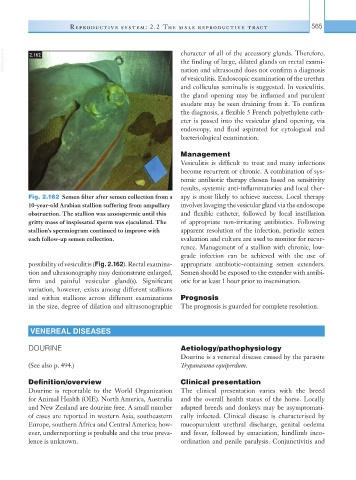
Fig. 2.162 Semen filter after semen collection from a apy is most likely to achieve success. Local therapy
10-year-old Arabian stallion suffering from ampullary involves lavaging the vesicular gland via the endoscope
obstruction. The stallion was azoospermic until this and flexible catheter, followed by local instillation
gritty mass of inspissated sperm was ejaculated. The of appropriate non-irritating antibiotics. Following
stallion’s spermiogram continued to improve with apparent resolution of the infection, periodic semen
each follow-up semen collection. evaluation and culture are used to monitor for recur-
rence. Management of a stallion with chronic, low-
grade infection can be achieved with the use of
possibility of vesiculitis (Fig. 2.162). Rectal examina- appropriate antibiotic-containing semen extenders.
tion and ultrasonography may demonstrate enlarged, Semen should be exposed to the extender with antibi-
firm and painful vesicular gland(s). Significant otic for at least 1 hour prior to insemination.
variation, however, exists among different stallions
and within stallions across different examinations Prognosis
in the size, degree of dilation and ultrasonographic The prognosis is guarded for complete resolution.
VENEREAL DISEASES
DOURINE Aetiology/pathophysiology
Dourine is a venereal disease caused by the parasite
(See also p. 494.) Trypanasoma equiperdum.
Definition/overview Clinical presentation
Dourine is reportable to the World Organization The clinical presentation varies with the breed
for Animal Health (OIE). North America, Australia and the overall health status of the horse. Locally
and New Zealand are dourine free. A small number adapted breeds and donkeys may be asymptomati-
of cases are reported in western Asia, southeastern cally infected. Clinical disease is characterised by
Europe, southern Africa and Central America; how- mucopurulent urethral discharge, genital oedema
ever, underreporting is probable and the true preva- and fever, followed by emaciation, hindlimb inco-
lence is unknown. ordination and penile paralysis. Conjunctivitis and