
Page 620 - Equine Clinical Medicine, Surgery and Reproduction, 2nd Edition
P. 620
Respir atory system: 3.1 Introduction 595
VetBooks.ir 3.6 3.7
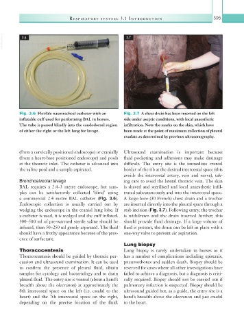
Fig. 3.6 Flexible nasotracheal catheter with an Fig. 3.7 A chest drain has been inserted on the left
inflatable cuff used for performing BAL in horses. side under aseptic conditions, with local anaesthetic
The tube is passed blindly into the caudodorsal region infiltration. Note the marks on the skin, which have
of either the right or the left lung for lavage. been made at the point of maximum collection of pleural
exudate as determined by previous ultrasonography.
(from a cervically positioned endoscope) or cranially Ultrasound examination is important because
(from a heart-base positioned endoscope) and pools fluid pocketing and adhesions may make drainage
at the thoracic inlet. The catheter is advanced into difficult. The entry site is the immediate cranial
the saline pool and a sample aspirated. border of the rib at the desired intercostal space (this
avoids the intercostal artery, vein and nerve), tak-
Bronchoalveolar lavage ing care to avoid the lateral thoracic vein. The skin
BAL requires a 2.4–3 metre endoscope, but sam- is shaved and sterilised and local anaesthetic infil-
ples can be satisfactorily collected ‘blind’ using trated subcutaneously and into the intercostal space.
a commercial 2.4 metre BAL catheter (Fig. 3.6). A large-bore (30 French) chest drain and a trochar
Endoscopic collection is usually carried out by are inserted directly into the pleural space through a
wedging the endoscope in the cranial lung lobe. If stab incision (Fig. 3.7). Following entry, the trochar
a catheter is used, it is wedged and the cuff inflated. is withdrawn and the drain inserted further; this
300–500 ml of pre-warmed sterile saline should be should provide fluid drainage. If a large volume of
infused, then 50–250 ml gently aspirated. The fluid fluid is present, the drain can be left in place with a
should have a frothy appearance because of the pres- one-way valve to prevent air aspiration.
ence of surfactant.
Lung biopsy
Thoracocentesis Lung biopsy is rarely undertaken in horses as it
Thoracocentesis should be guided by thoracic per- has a number of complications including epistaxis,
cussion and ultrasound examination. It can be used pneumothorax and sudden death. Biopsy should be
to confirm the presence of pleural fluid, obtain reserved for cases where all other investigations have
samples for cytology and bacteriology and to drain failed to achieve a diagnosis, but a diagnosis is criti-
pleural fluid. The entry site is ventral (about a hand’s cally required. Biopsy should not be carried out if
breadth above the olecranon) at approximately the pulmonary infection is suspected. Biopsy should be
8th intercostal space on the left (i.e. caudal to the ultrasound guided but, as a guide, the entry site is a
heart) and the 7th intercostal space on the right, hand’s breadth above the olecranon and just caudal
depending on the precise location of the fluid. to the heart.