
Page 73 - Equine Clinical Medicine, Surgery and Reproduction, 2nd Edition
P. 73
48 CHAPTER 1
VetBooks.ir 1.84
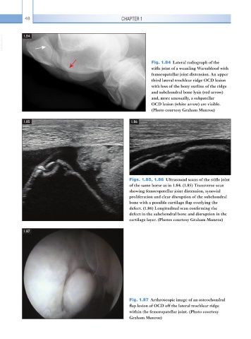
Fig. 1.84 Lateral radiograph of the
stifle joint of a weanling Warmblood with
femoropatellar joint distension. An upper
third lateral trochlear ridge OCD lesion
with loss of the bony outline of the ridge
and subchondral bone lysis (red arrow)
and, more unusually, a subpatellar
OCD lesion (white arrow) are visible.
(Photo courtesy Graham Munroe)
1.85 1.86
Figs. 1.85, 1.86 Ultrasound scans of the stifle joint
of the same horse as in 1.84. (1.85) Transverse scan
showing femoropatellar joint distension, synovial
proliferation and clear disruption of the subchondral
bone with a possible cartilage flap overlying the
defect. (1.86) Longitudinal scan confirming the
defect in the subchondral bone and disruption in the
cartilage layer. (Photos courtesy Graham Munroe)
1.87
Fig. 1.87 Arthroscopic image of an osteochondral
flap lesion of OCD off the lateral trochlear ridge
within the femoropatellar joint. (Photo courtesy
Graham Munroe)