
Page 731 - Equine Clinical Medicine, Surgery and Reproduction, 2nd Edition
P. 731
706 CHAPTER 3
MISCELLANEOUS CONDITIONS
VetBooks.ir ASPIRATION PNEUMONIA 3.165
Definition/overview
Aspiration is a potential cause of serious pneumonia.
It may occur in adult horses and foals for a variety
of reasons.
Aetiology/pathophysiology
There are many potential causes of aspiration. In
neonatal foals, aspiration of milk may occur because
of congenital abnormalities, weakness secondary
to other illness or improper supplemental (bottle
or nasogastric tube) feeding. Inadvertent drench-
ing or passing a nasogastric tube into the lungs and
depositing fluids, pharmaceutical products or other
substances into the lower airways is a common cause
of aspiration in adult horses. Aspiration of saliva
and feed material is also a common complication of
oesophageal obstruction (choke) and may also occur
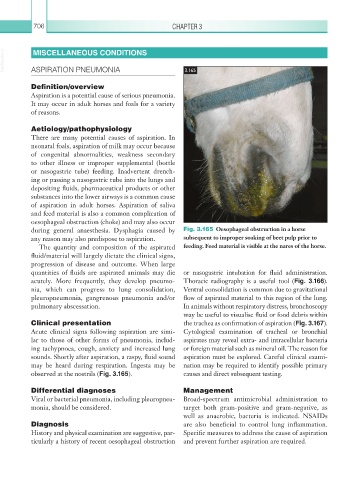
during general anaesthesia. Dysphagia caused by Fig. 3.165 Oesophageal obstruction in a horse
any reason may also predispose to aspiration. subsequent to improper soaking of beet pulp prior to
The quantity and composition of the aspirated feeding. Feed material is visible at the nares of the horse.
fluid/material will largely dictate the clinical signs,
progression of disease and outcome. When large
quantities of fluids are aspirated animals may die or nasogastric intubation for fluid administration.
acutely. More frequently, they develop pneumo- Thoracic radiography is a useful tool (Fig. 3.166).
nia, which can progress to lung consolidation, Ventral consolidation is common due to gravitational
pleuropneumonia, gangrenous pneumonia and/or flow of aspirated material to this region of the lung.
pulmonary abscessation. In animals without respiratory distress, bronchoscopy
may be useful to visualise fluid or food debris within
Clinical presentation the trachea as confirmation of aspiration (Fig. 3.167).
Acute clinical signs following aspiration are simi- Cytological examination of tracheal or bronchial
lar to those of other forms of pneumonia, includ- aspirates may reveal extra- and intracellular bacteria
ing tachypnoea, cough, anxiety and increased lung or foreign material such as mineral oil. The reason for
sounds. Shortly after aspiration, a raspy, fluid sound aspiration must be explored. Careful clinical exami-
may be heard during respiration. Ingesta may be nation may be required to identify possible primary
observed at the nostrils (Fig. 3.165). causes and direct subsequent testing.
Differential diagnoses Management
Viral or bacterial pneumonia, including pleuropneu- Broad-spectrum antimicrobial administration to
monia, should be considered. target both gram-positive and gram-negative, as
well as anaerobic, bacteria is indicated. NSAIDs
Diagnosis are also beneficial to control lung inflammation.
History and physical examination are suggestive, par- Specific measures to address the cause of aspiration
ticularly a history of recent oesophageal obstruction and prevent further aspiration are required.