
Page 742 - Equine Clinical Medicine, Surgery and Reproduction, 2nd Edition
P. 742
Respir atory system: 3.4 Medical conditions of the lower respir atory tr act 717
VetBooks.ir Prognosis effort with nasal flare and extended head and neck.
Foals are often febrile. Auscultation of lung fields
The prognosis is generally poor, although it may
depend on the type of tumour. By the time the
wheezes, apparent in areas where sufficient air move-
tumour is clinically evident and is subsequently reveals reduced air movement with crackles and
diagnosed, lesions are frequently advanced and the ment remains. Cough and nasal discharge are vari-
animal has developed systemic complications. ably present.
ACUTE BRONCHOINTERSTITIAL Differential diagnosis
PNEUMONIA OF FOALS Differential diagnosis includes bacterial pneumonia or
pleuropneumonia, fungal pneumonia and Pneumocystis
Definition/overview jiroveci infection of immunocompromised foals.
This is a rare condition in foals less than 10 months
of age, which are affected by the acute onset of Diagnosis
severe interstitial lung disease. Respiratory distress Laboratory findings of affected foals include neutro-
is apparent and the disease progresses rapidly. The philia, with or without a left shift, and elevated fibrin-
prognosis is poor, although foals surviving past the ogen. If blood gas analysis is available, hypoxaemia is
first 7–10 days are likely to recover. present. Azotaemia may reflect dehydration second-
ary to the reduced intake of fluids. Ultrasonography
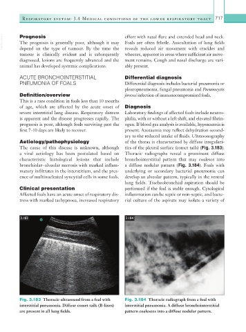
Aetiology/pathophysiology of the thorax is characterised by diffuse irregulari-
The cause of this disease is unknown, although ties of the pleural surface (comet tails) (Fig. 3.183).
a viral aetiology has been postulated based on Thoracic radiographs reveal a prominent diffuse
characteristic histological lesions that include bronchointerstitial pattern that may coalesce into
bronchiolar–alveolar necrosis with marked inflam- a diffuse nodular pattern (Fig. 3.184). Foals with
matory infiltrates in the interstitium, and the pres- underlying or secondary bacterial pneumonia can
ence of multinucleated syncytial cells in some foals. develop an alveolar pattern, typically in the ventral
lung fields. Tracheobronchial aspiration should be
Clinical presentation performed if the foal is stable enough. Cytological
Affected foals have an acute onset of respiratory dis- inflammation can be septic or non-septic, and bacte-
tress with marked tachypnoea, increased respiratory rial culture of the aspirate may isolate a variety of
3.183 3.184
Fig. 3.183 Thoracic ultrasound from a foal with Fig. 3.184 Thoracic radiograph from a foal with
interstitial pneumonia. Diffuse comet tails (B-lines) interstitial pneumonia. A diffuse bronchointerstitial
are present in all lung fields. pattern coalesces into a diffuse nodular pattern.