
Page 753 - Equine Clinical Medicine, Surgery and Reproduction, 2nd Edition
P. 753
728 CHAPTER 4
VetBooks.ir 4.11 4.12
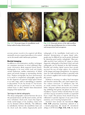
Fig. 4.11 Oroscopic image of a mandibular tooth Fig. 4.12 Oroscopic picture of the right maxillary
being explored using a fissure probe. arcade showing malalignment due to overcrowding
and interproximal food entrapment.
accurate picture records to be acquired and allows radiographs of the mandibular cheek teeth to be
minimally-invasive surgical procedures to be carried obtained. Interpretation of equine dental radio-
out in the mouth under endoscopic guidance. graphs may be difficult but can be greatly facilitated
by obtaining good quality radiographs. Most por-
Dental imaging table machines have sufficient kV output to obtain
In addition to oral examination, ancillary techniques diagnostic radiographs of the horse’s head, and such
are sometimes necessary to reveal additional diag- radiographs can be obtained in the field situation.
nostic information about suspected dental disease. Portable and wireless digital systems have greatly
Radiography is the most widely used, and despite increased the ease of image capture, the resolution
certain limitations, enables examination of dental of the images and image manipulation. An exposure
apices and reveals changes to surrounding alveolar chart for each individual machine is provided with
bone. Nuclear scintigraphy has been demonstrated the software supplied with modern digital radiogra-
to be useful to complement radiography, especially phy systems
for the diagnosis of suspected dental apical disease Sedation is necessary to reduce head movement
at an early stage, such as remodelling in response and therefore motion blur, and a head support fur-
to abscessation. In the last decade CT has become ther reduces movement. A radiolucent rope hal-
available and can be performed in the conscious ter avoids artefacts from the buckles of a normal
sedated horse to allow detailed three-dimensional halter. Adequate radiation protection and monitor-
imaging of the equine head. ing, including lead gowns and gloves or sleeves, is
mandatory. Cassette holders should always be used
Technique for dental radiographs and these may be mounted on handles or suspended
The sharp radiographic contrast between the air in from a stand. The primary beam should always be
the paranasal sinuses and the nasal cavities, and the collimated to prevent exposure of any parts of the
more radiodense cheek teeth, means that diagnos- assistants, even if wearing lead protection.
tically useful images of the maxillary cheek teeth Standard views include the laterolateral (Figs.
can be obtained without difficulty. The difference 4.13, 4.14) projection (sometimes referred to as
in radiodensity between the mandibular bone and a lateral projection), the lateral 30° dorsolateral
the cheek teeth also enables diagnostically useful oblique (L30°D-LO) (Figs. 4.15, 4.16), the lateral