
Page 827 - Equine Clinical Medicine, Surgery and Reproduction, 2nd Edition
P. 827
802 CHAPTER 4
VetBooks.ir also induce the formation of abdominal adhesion in Differential diagnosis
Any clinical condition that may induce acute non-
horses. Foals under the age of 30 days are reported
to be substantially more susceptible to postoperative
tion, recurrent colic and poor body condition should
adhesion formation, but this is largely dependent on strangulating or strangulating intestinal obstruc-
the degree of underlying systemic illness. be considered.
Abdominal adhesions form at a peritoneal injury
site as a result of an imbalance between fibrin depo- Diagnosis
sition and fibrinolysis. Inflammation and ischaemia Abdominal adhesions are diagnosed most frequently
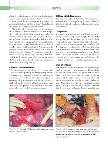
increase fibrin deposition and decrease fibrinoly- during exploratory laparotomy (Figs. 4.127, 4.128).
sis. Adhesions begin to form within 48 hours of an Rarely, they can be palpated p/r as a tight band
injury as a fibrin cover appears on the injured serosal running from one viscus to either the body wall or
bed. Adhesions are well formed by 5–7 days, but are another viscus. Ultrasonography may be helpful with
usually not irreversible until after 7 days, when the the diagnosis of abdominal adhesions. Intestinal
collagen content increases to a level that cannot be adhesions should be suspected in horses with a his-
inherently broken down. Permanent fibrous adhe- tory of previous coeliotomy and appropriate clinical
sions are usually formed by 7–14 days. Extensive signs. The ventral midline should be palpated for
well-defined adhesions are often covered by meso- evidence of previous surgery, particularly when the
thelium and contain blood vessels and connective complete health history of the horse is unknown.
tissue fibres, including elastin.
Management
Clinical presentation If the signs of colic resulting from adhesions are mild,
The most common clinical sign is recurrent colic. medical treatment can be attempted. This includes
Acute non-strangulating or strangulating intesti- the use of enteral fluids, analgesics and modified
nal obstruction can occur in horses with abdominal diets. Low-residue feeds, such as completely pelleted
adhesions. Poor body condition and an inability to feeds or alfalfa cubes, may pass through the small
consume a high-roughage diet may also be associ- intestine more easily and reduce the risk of obstruc-
ated with mild restrictive adhesions. Clinical signs tion. After several weeks on a modified feeding
associated with postoperative abdominal adhesions regime, it may be possible to re-institute a normal
are usually observed 1–4 weeks after surgery. diet if the fibrous adhesions have remodelled and
4.127 4.128
Fig. 4.127 Example of fibrinous adhesions that cover Fig. 4.128 Example of a focal mesentery-to-intestine
injured serosal surfaces. adhesion. Note the ischaemic (pale) area on the intestinal
surface that most likely induced the formation of this
adhesion (arrow).