
Page 201 - Anatomy and Physiology of Farm Animals, 8th Edition
P. 201
186 / Anatomy and Physiology of Farm Animals
Action potentials
VetBooks.ir 20
Membrane potential (mV) –20
–60
5 15 25
Time (s)
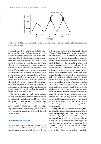
Figure 9-10. Slow‐wave electrical activity in smooth muscle with action potentials occurring at the
peak of slow waves.
contractions, but action potentials may a branching network of terminal fibrils.
occur at the peak of these waves, and the These fibrils have varicosities (beadlike
action potentials are associated with con- enlargements) at intervals along their
tractions (Fig. 9‐10). Because action poten- axons. When action potentials depolarize
tials and contractions are seen only at the them, the transmitter substance is released
peak of the slow waves, the rate at which and diffuses to the smooth muscle cell
slow waves develop determines the rate at membranes of several cells, where stimu-
which smooth muscle contractions can lation occurs. In multiunit smooth muscle,
occur. The precise origin of the slow waves a branch of an autonomic nerve inner-
is uncertain, but a variety of stimuli, such vates each muscle fiber. This provides
as hormones, neurotransmitters, and the more direct neural control of each muscle
local chemical environment, can deter- cell, but the junction between neuron and
mine whether action potentials occur at muscle is less highly structured than the
the peak of the slow waves. The relation- neuromuscular junction of skeletal muscle.
ship between slow‐wave activity and action For both types of smooth muscle, the
potentials is important in the regulation of innervation is usually dual; that is, both
gastrointestinal motility and is discussed in divisions of the autonomic nervous sys-
more detail in Chapter 21. tem innervate smooth muscle. Important
Action potentials spread across groups exceptions are blood vessels (arteries,
of single‐unit smooth muscle fibers because arterioles, and veins), which have pre-
of gap junctions between the fibers (where dominantly sympathetic innervation, and
the plasma membranes of adjacent cells in the skin, where the pilomotor fibers
touch). Thus, single‐unit smooth muscle and sweat glands receive only sympathetic
cells can be linked electrically while remain- innervation.
ing independent chemically (no secretion Acetylcholine is released from the par-
of transmitter substance from cell to cell asympathetic postganglionic nerve fibers
is required). and norepinephrine from the sympa-
thetic postganglionic fibers. The response
of smooth muscle (contraction or relaxa-
Autonomic Innervation tion) to these neuromediators depends
on the type of autonomic receptor (see
In visceral or single‐unit smooth muscle, the Tables 11‐1 and 11‐2) on the smooth mus-
fibers of the autonomic nervous system cle and the intracellular events initiated by
travel between the smooth muscle cells in the binding of the neuromediators to their