
Page 1081 - Clinical Small Animal Internal Medicine
P. 1081
110 Protozoal and Protozoa‐Like Infections 1019
susceptibilities, treatment with one of these drug options such as amphotericin B, ketoconazole, and miltefosine
VetBooks.ir should be replaced by another if it is not successful in for deep organ infection.
effectively relieving clinical signs. Furthermore, co‐infec-
tions should be diagnosed and treated and sanitary man-
agement with quaternary ammonium disinfectants Trichomoniasis
should be instituted to prevent reinfection. Trichomonas spp. are flagellated protozoa that belong to
the order Trichomonadida, reproduce by binary fission,
and do not produce cysts. Cats and more rarely dogs can
Prognosis be infected with Tritrichomonas foetus which is trans-
The prognosis of treated dogs and cats is usually good mitted by the direct fecal–oral route and causes diarrhea
and clinical signs are ameliorated, although some ani- [31,32]. Tritrichomonas foetus survives in feces and
mals could suffer from persistent or recurrent infection. humid environment outside the animal’s body for up to a
few days. The parasite has been reported in numerous
countries and has a worldwide distribution, with infection
Prevention rates of up to 32% in cat populations from different coun-
Disinfection of kennels, cages, and the animal’s direct tries in Europe [32]. Subclinical infection with T. foetus is
environment, boiling or filtering drinking water and common and there is not a direct relationship between
removal of feces are important for controlling the spread infection and clinical signs. Infection is more common in
of infection among animals that live in close proximity. cats from multicat settings and breeding catteries
Bathing of dogs and cats in infected kennels may decrease and clinical signs are more common in young cats under
transmission of giardiasis between individual animals. 12 months of age.
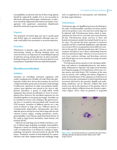
Tritrichomonas foetus attaches to the intestinal epithe-
lium and induces a lymphoplamsmacytic and neutro-
Miscellaneous Infections philic inflammatory response with large bowel diarrhea
and frequent passage of soft liquid feces which may con-
tain mucus and fresh blood [33]. Irritation of the anus
Amebiasis
may be present with swelling and edema. Diagnosis is
Amoebas are unicellular protozoal organisms with made by identification of the organism in fresh feces by
motile cytoplasm and a flexible cell wall. Molecular phy- direct microscopic examination in which typical forward
logenetic studies have shown that amoebas do not form movement directed by the parasite’s flagella and undu-
a single taxonomic group and are found in many lineages lating membrane can be detected. Stained thin fecal
of eukaryotic organisms. In older nomenclature classifi- smears may aid in the detection of the parasite with its
cations, most amoebas were placed in the class or sub- typical form which is different from the Giardia tropho-
phylum Sarcodina, a group of single‐celled motile zoites (Figure 110.5), when its presence is suspected
organisms that possess pseudopods or move by proto-
plasmic flow. However, the amoeboid organisms are not
classified together in one group any longer. Amoebas can
be classified as enteric and residing in the gastrointesti-
nal tract or nonenteric and associated with visceral and
CNS invasion. Amoebas of different species are a rare
cause of disease in dogs and cats. Different species of
Acanthamoeba, Balamuthia, Hartmannella, and other
amoebic genera and species may infect dogs and cats and
colonize various organs, causing pneumonia, menin-
goencephalitis, renal disease, gastrointestinal disease of
the small or large bowel, dermatitis, nasal disease, and
keratitis [29,30].
Affected cats and dogs may be febrile and show a vari-
ety of clinical signs including neurologic abnormalities.
Diagnosis is based on finding amoebae in excretions as
cysts or trophozoites, or in tissues by cytology or histo-
pathology and genetic characterization by specific PCR Figure 110.5 Tritrichomonas foetus in a stained fecal smear from a
assays. Treatment consists of metronidazole for enteric naturally infected cat (May Grunwald–Giemsa stain 1000×
infection and other antiprotozoal and antifungal drugs magnification).