
Page 1326 - Clinical Small Animal Internal Medicine
P. 1326
1264 Section 11 Oncologic Disease
growth rate of these tumors. If intervention is required, Most tumors are benign and detected in older dogs
VetBooks.ir lamellar keratectomy/sclerectomy with graft placement, (7 years of age or older) although more rapidly growing
pigmented iridal masses have been described in young
cryosurgery or laser photocoagulation may be curative
or reduce the size of the mass such that it no longer
invasive but distant metastasis (usually hematogenously)
threatens vision for the remainder of the animal’s life. dogs. Malignant intraocular melanomas may be locally
Cryotherapy or beta‐irradiation may also substantially is rare.
reduce the risk of recurrence following local excision. Ocular melanosis of cairn terriers is a distinct clini-
Enucleation is curative and indicated if the mass has cal entity that resembles a diffuse and benign form of
entered the eye and there is ocular discomfort. uveal melanoma. It is a bilateral disorder in which the
iris becomes thickened and more darkly pigmented,
followed by release of pigment into the aqueous and
Intraocular Tumors pigment deposition in the sclera/episclera. Secondary
glaucoma is common. Some cairn terriers may have
Primary Tumors overt uveal melanocytic tumors, but in most dogs the
pigmented cells are nonneoplastic (Figure 138.2).
Primary intraocular tumors occur more frequently than
metastatic intraocular neoplasms in dogs and cats. They
most commonly originate from the uvea, and their etiol- Choroidal Melanocytoma/Melanoma
ogy is poorly understood. These rare melanocytic tumors generally originate
from the peripapillary choroid and appear as well‐
Canine Anterior Uveal Melanocytoma/Melanoma delineated, raised, subretinal pigmented masses. They
Intraocular melanocytomas arising from the iris or ciliary may remain virtually static for many years, although
body comprise the majority of canine intraocular tumors. others may extend into the overlying retina, sclera,
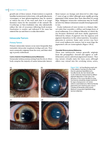
(a) (b) Figure 138.2 (a) Free‐floating uveal cysts
in the anterior chamber of a dog. Note
that the spherical, fluid‐filled cysts
transilluminate. (b) Cairn terrier with
ocular melanosis characterized by diffuse
pigment deposition in the sclera and
episclera. (c) Iris nevus (freckle) in a dog.
Such lesions should be closely monitored
for signs of progression. (d) Canine
anterior uveal melanoma. Note the heavily
pigmented and raised iris mass. Source:
Courtesy of University of Wisconsin‐
Madison Comparative Ophthalmology
Service Collection.
(c) (d)