
Page 693 - Clinical Small Animal Internal Medicine
P. 693
61 Imaging in Hepatobiliary Disease 661
(a) (b)
VetBooks.ir
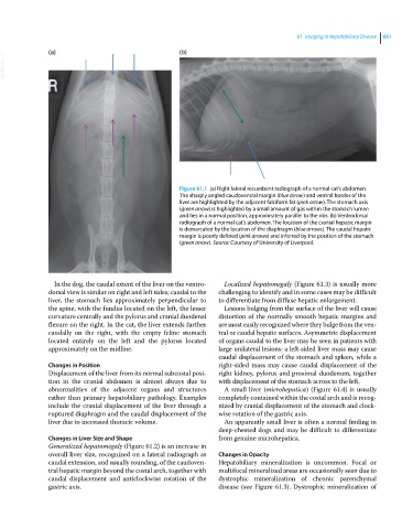
Figure 61.1 (a) Right lateral recumbent radiograph of a normal cat’s abdomen.
The sharply angled caudoventral margin (blue arrow) and ventral border of the
liver are highlighted by the adjacent falciform fat (pink arrow). The stomach axis
(green arrow) is highlighted by a small amount of gas within the stomach lumen
and lies in a normal position, approximately parallel to the ribs. (b) Ventrodorsal
radiograph of a normal cat’s abdomen. The location of the cranial hepatic margin
is demarcated by the location of the diaphragm (blue arrows). The caudal hepatic
margin is poorly defined (pink arrows) and inferred by the position of the stomach
(green arrow). Source: Courtesy of University of Liverpool.
In the dog, the caudal extent of the liver on the ventro- Localized hepatomegaly (Figure 61.3) is usually more
dorsal view is similar on right and left sides; caudal to the challenging to identify and in some cases may be difficult
liver, the stomach lies approximately perpendicular to to differentiate from diffuse hepatic enlargement.
the spine, with the fundus located on the left, the lesser Lesions bulging from the surface of the liver will cause
curvature centrally and the pylorus and cranial duodenal distortion of the normally smooth hepatic margins and
flexure on the right. In the cat, the liver extends further are most easily recognized where they bulge from the ven-
caudally on the right, with the empty feline stomach tral or caudal hepatic surfaces. Asymmetric displacement
located entirely on the left and the pylorus located of organs caudal to the liver may be seen in patients with
approximately on the midline. large unilateral lesions: a left‐sided liver mass may cause
caudal displacement of the stomach and spleen, while a
Changes in Position right‐sided mass may cause caudal displacement of the
Displacement of the liver from its normal subcostal posi- right kidney, pylorus and proximal duodenum, together
tion in the cranial abdomen is almost always due to with displacement of the stomach across to the left.
abnormalities of the adjacent organs and structures A small liver (microhepatica) (Figure 61.4) is usually
rather than primary hepatobiliary pathology. Examples completely contained within the costal arch and is recog-
include the cranial displacement of the liver through a nized by cranial displacement of the stomach and clock-
ruptured diaphragm and the caudal displacement of the wise rotation of the gastric axis.
liver due to increased thoracic volume. An apparently small liver is often a normal finding in
deep‐chested dogs and may be difficult to differentiate
Changes in Liver Size and Shape from genuine microhepatica.
Generalized hepatomegaly (Figure 61.2) is an increase in
overall liver size, recognized on a lateral radiograph as Changes in Opacity
caudal extension, and usually rounding, of the caudoven- Hepatobiliary mineralization is uncommon. Focal or
tral hepatic margin beyond the costal arch, together with multifocal mineralized areas are occasionally seen due to
caudal displacement and anticlockwise rotation of the dystrophic mineralization of chronic parenchymal
gastric axis. disease (see Figure 61.3). Dystrophic mineralization of