
Page 113 - BSAVA Manual of Canine and Feline Head, Neck and Thoracic Surgery, 2nd Edition
P. 113
BSAVA Manual of Canine and Feline Head, Neck and Thoracic Surgery
of subcutaneous emphysema alone. If a cervical bite wound addition to the absence of normal laryngeal function, may
is suspected, radiographs must be obtained of the neck suggest that placement of a temporary tracheostomy tube
VetBooks.ir or mediastinal emphysema in all animals with airway injury patient basis. The cervical wound must be thoroughly
and thoracic cavity. Radiography revealed subcutaneous
is indicated, but this must be decided on an individual
lavaged and can be closed over a drain once ‘healthy’. The
following cervical bite wounds in one study of 56 cases
(Jordan et al., 2013). Trach eoscopy may also be used to
location as active suction drains could cause damage to
evaluate the presence, extent and location of tracheal injury. use of a Penrose drain may be most appropriate at this
All animals with cervical bite wounds should undergo important cervical neurovascular structures. If use of a
ventral midline neck exploration for appropriate wound closed suction drain is indicated then the most appropriate
management, particularly if emphysema has been detected drain to select is a Jackson Pratt drain, which has a more
indicating that airway repair may be required. Given the consistent low level of suction than other drain types
location of the trauma, injury to other important structures, (Halfacree et al., 2009).
such as neurovascular bundles, the spine, pharynx or Iatrogenic tracheal tears can occur acutely following
oesophagus, may have occurred. endotracheal intubation, due to tube cuff overinflation,
Following identification of an injury to the trachea, the frequent changes in patient position without disconnect-
tissue must be assessed for viability and debridement ing the endotracheal tube (e.g. during anaesthesia for
performed where necessary. Primary repair may be dental prophylaxis) or use of an overlong stylet. Pressure
pos sible for a small defect (Figure 8.3); a fasciomuscular necrosis of the trachea following prolonged use of a low-
patch, such as sternohyoid muscle (Jordan et al., 2013), volume high-pressure cuff can also cause gradual loss of
can be used to augment the repair if necessary. Where a tracheal integrity, therefore resulting in a delay in presen-
section of trachea is non-viable, a tracheal resection and tation of clinical signs. The clinical signs observed include
anastomosis should be performed. In all cases, great care coughing, dyspnoea and the development of subcuta-
must be taken to preserve the segmental blood supply to neous emphysema. Radiography of the neck and thorax
the trachea and to avoid injury to the recurrent laryngeal reveals subcutaneous emphysema and pneumomedia-
nerves running in close approximation to the trachea. stinum (Figure 8.4). The majority of cats can be treated
Airway examination should be conducted to assess laryn- conservatively with strict cage rest; however, surgery
geal function both before and after the surgery. If the site may be required (Mitchell et al., 2000). The trachea is
of tracheal injury is evident, it is prudent to select a cuffed approached via a ventral midline cervical approach and
endotracheal tube that can be passed beyond the site of the lesion in the dorsal tracheal ligament is debrided
perforation and therefore allow controlled management and repaired. The use of high-volume low-pressure cuffs,
of anaesthesia and ventilation if required. Significant air- rather than low-volume high-pressure cuffs, is recom-
way swelling may be present in some patients and this, in mended to minimize the risk of this injury.
(b)
(a)
(c) (d)
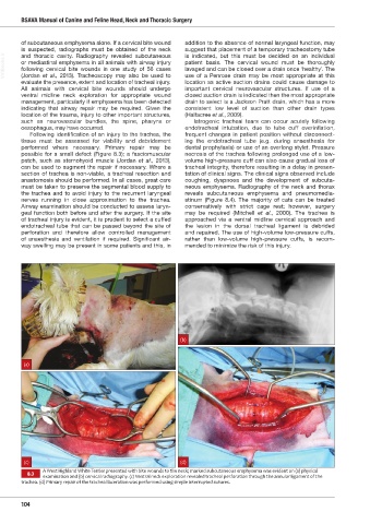
A West Highland White Terrier presented with bite wounds to the neck; marked subcutaneous emphysema was evident on (a) physical
8.3
examination and (b) cervical radiography. (c) Ventral neck exploration revealed tracheal perforation through the annular ligament of the
trachea. (d) Primary repair of the tracheal laceration was performed using simple interrupted sutures.
104
Ch08 HNT.indd 104 31/08/2018 11:32