
Page 119 - BSAVA Manual of Canine and Feline Head, Neck and Thoracic Surgery, 2nd Edition
P. 119
BSAVA Manual of Canine and Feline Head, Neck and Thoracic Surgery
aspect of the trachea, tracking laterally and then ventro- to ensure it has not been sutured; alternatively, the cuff is
laterally as it courses caudally towards the thoracic inlet located prior to suture placement and avoided.
VetBooks.ir located within the carotid sheath (Fossum, 2002). Damage ization to reduce catastrophic perioperative complications
One report advocates concurrent left arytenoid lateral-
(Figure 8.11b); the right recurrent laryngeal nerve may be
associated with iatrogenic laryngeal paralysis (White,
to either of these important structures during the procedure
l
can lead to tracheal necrosis or laryngeal para ysis, respec-
tively. Excessive dissection around the trachea should be 1995), but the authors of this chapter do not routinely per-
form this additional procedure. Repeat tracheoscopy is
avoided by gentle blunt dissection and fenestration only performed following the procedure to confirm appropriate
where the ring is to be placed. luminal patency following placement of tracheal rings.
A pair of curved haemostats or right-angled Mixter During recovery and spontaneous breathing, one can
forceps facilitates passage of the ring around the collapsed attempt to evaluate for laryngeal paralysis although
trachea, and 1.5 metric (4/0 USP) or 2 metric (3/0 USP) assessment can be difficult following general anesthesia.
i
monofilament non-absorbable suture material is passed Mon toring would be recommended before surgical treat-
through the ring and trachea. It is imperative that at least ment would be indicated.
one suture engages the dorsal tracheal membrane. Care is
taken to avoid the endotracheal tube cuff during passage of Results: The largest retrospective study evaluating the use
the suture into the tracheal lumen. Temporarily leaving one of extraluminal polypropylene ring prostheses for tracheal
of the ventral sutures long, for use as a stay suture, can collapse (Buback et al., 1996) reported a 5% perioperative
facilitate cranial traction to increase exposure to part of the mortality rate, a 37% rate of immediate postoperative
intrathoracic trachea without extension of the incision into complications (24% coughing, 16% dyspnoea, and 11%
a thoracotomy. Care must be taken to avoid penetration of incidence of laryngeal paralysis) and a 19% incidence of
the pleural cavity and subsequent pneumothorax. Rings permanent tracheostomy (more than half of which were
are placed approximately 5 mm apart (Figure 8.11cd). The performed within 24 hours of surgery). Only 10% of the 90
endotracheal tube is moved gently after each ring is placed dogs in this study had evidence of intrathoracic tracheal
collapse for which the perioperative morbidity was exces-
sive enough to recommend avoiding surgery. For those
animals that recovered favourably, the median survival time
was approximately 2 years; half of these animals died of
causes unrelated to the respiratory system. Age at the time
of surgery was the only prognostic factor identified:
animals younger than 6 years had more severe tracheal
collapse but a better prognosis.
(a) Some smaller studies report more favourable results,
such as a 4% complication rate and 75% success rate
when concurrent left arytenoid lateralization is performed
(White, 1995). More recently, two other studies have
reported improved outcomes in terms of prolonged survival
times (>2500 days for cervical collapse alone) and a
reduced need for postoperative medications; however, rates
of laryngeal paralysis and other respiratory complications
were still high (Becker et al., 2012; Chisnell and Pardo,
(b) 2015). Tinga et al. (2015) demonstrated similar major compli-
cations for dogs with stents or extraluminal rings, and no
difference in median survival times when corrected for age.
In general, it appears that animals with concurrent
cardiac or respiratory disease or mainstem bronchial col-
lapse may have a worse prognosis. It is clear that careful
patient selection and long discussions with the animal s
owners are important to explain potential complications
and expectations.
Intraluminal devices
(c)
Interventional radiology involves the use of imaging modal-
ities, such as fluoroscopy, to gain access to structures
to administer materials or devices for thera peutic reasons.
Tracheal stenting, the minimally invasive, through-the-
mouth placement of a stent (support) within the lumen of
the trachea, has been investigated. Migration of balloon-
expandable stents led researchers to evaluate various
types of self-expanding stents made of stainless steel or
(d) nitinol (a nickel–titanium alloy) (Radlinsky et al., 1997).
Stenting provides a rapid, minimally invasive option that
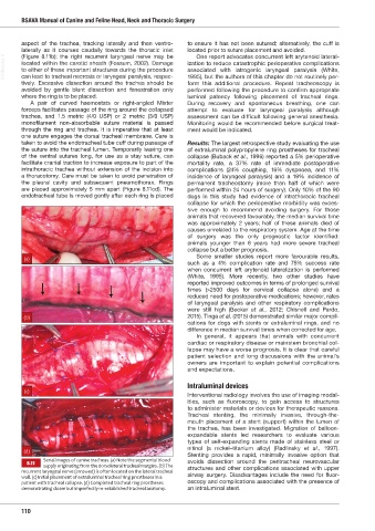
erial images of canine tracheas. a ote the segmental blood
8.11 avoids dissection around the peritracheal neurovascular
supply originating from the dorsolateral tracheal margins. (b) The
recurrent laryngeal nerve (arrowed) is often located on the lateral tracheal structures and other complications associated with upper
wall. (c) Initial placement of extraluminal tracheal ring prostheses in a airway surgery. Disadvantages include the need for fluor-
patient with tracheal collapse. (d) Completed tracheal ring prostheses oscopy and complications associated with the presence of
demonstrating closer but imperfectly re-established tracheal anatomy. an intraluminal stent.
110
Ch08 HNT.indd 110 31/08/2018 11:32