
Page 155 - BSAVA Manual of Canine and Feline Head, Neck and Thoracic Surgery, 2nd Edition
P. 155
BSAVA Manual of Canine and Feline Head, Neck and Thoracic Surgery
VetBooks.ir
(a) (b) (c)
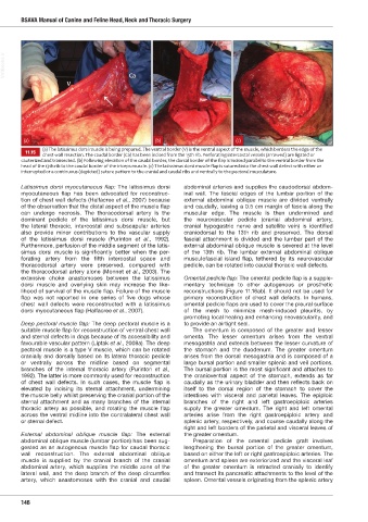
(a) The latissimus dorsi muscle is being prepared. The ventral border (V) is the ventral aspect of the muscle, which borders the edge of the
11.15 chest wall resection. The caudal border (Ca) has been incised from the 13th rib. Perforating intercostal vessels (arrowed) are ligated or
cauteri ed and transected. b Follo ing elevation of the caudal border the dorsal border of the flap is incised parallel to the ventral border from the
head of the 13th rib to the caudal border of the triceps muscle. c The latissimus dorsi muscle flap is sutured into the chest all defect ith either an
interrupted or a continuous (depicted) suture pattern to the cranial and caudal ribs and ventrally to the pectoral musculature.
i
Latissimus dorsi myocutaneous flap: The latissimus dorsi abdom nal arteries and supplies the caudodorsal abdom-
myocutaneous flap has been advocated for reconstruc- inal wall. The fascial edges of the lumbar portion of the
tion of chest wall defects (Halfacree et al., 2007) because external abdominal oblique muscle are divided ventrally
of the observation that the distal aspect of the muscle flap and caudally, leaving a 0.5 cm margin of fascia along the
can undergo necrosis. The thoracodorsal artery is the muscular edge. The muscle is then undermined and
dominant pedicle of the latissimus dorsi muscle, but the neurovascular pedicle (cranial abdominal artery,
the lateral thoracic, intercostal and subscapular arteries cranial hypogastric nerve and satellite vein) is identified
also provide minor contributions to the vascular supply craniodorsal to the 13th rib and preserved. The dorsal
of the latissimus dorsi muscle (Purinton et al., 1992). fascial attachment is divided and the lumbar part of the
Furthermore, perfusion of the middle segment of the latis- external abdominal oblique muscle is severed at the level
simus dorsi muscle is significantly better when the per- of the 13th rib. The lumbar external abdominal oblique
forating artery from the fifth intercostal space and musculofascial island flap, tethered by its neurovascular
thoracodorsal artery were preserved, compared with pedicle, can be rotated into caudal thoracic wall defects.
the thoracodorsal artery alone (Monnet et al., 2003). The
extensive choke anastomoses between the latissimus Omental pedicle flap: The omental pedicle flap is a supple-
dorsi muscle and overlying skin may increase the like- mentary technique to other autogenous or prosthetic
lihood of survival of the muscle flap. Failure of the muscle reconstructions (Figure 11.16ab). It should not be used for
flap was not reported in one series of five dogs whose primary reconstruction of chest wall defects. In humans,
chest wall defects were reconstructed with a latissimus omental pedicle flaps are used to cover the pleural surface
dorsi myocutaneous flap (Halfacree et al., 2007). of the mesh to minimize mesh-induced pleuritis, by
promoting local healing and enhancing neovascularity, and
Deep pectoral muscle flap: The deep pectoral muscle is a to provide an airtight seal.
suitable muscle flap for reconstruction of ventral chest wall The omentum is composed of the greater and lesser
and sternal defects in dogs because of its accessibility and omenta. The lesser omentum arises from the ventral
favourable vascular pattern (Liptak et al., 2008a). The deep mesogastria and extends between the lesser curvature of
pectoral muscle is a type V muscle, which can be rotated the stomach and the duodenum. The greater omentum
cranially and dorsally based on its lateral thoracic pedicle arises from the dorsal mesogastria and is composed of a
or ventrally across the midline based on segmental large bursal portion and smaller splenic and veil portions.
branches of the internal thoracic artery (Purinton et al., The bursal portion is the most significant and attaches to
1992). The latter is more commonly used for reconstruction the cranioventral aspect of the stomach, extends as far
of chest wall defects. In such cases, the muscle flap is caudally as the urinary bladder and then reflects back on
elevated by incising its sternal attachment, undermining itself to the dorsal region of the stomach to cover the
the muscle belly whilst preserving the cranial portion of the intestines with visceral and parietal leaves. The epiploic
sternal attachment and as many branches of the internal branches of the right and left gastroepiploic arteries
thoracic artery as possible, and rotating the muscle flap supply the greater omentum. The right and left omental
across the ventral midline into the contralateral chest wall arteries arise from the right gastroepiploic artery and
or sternal defect. splenic artery, respectively, and course caudally along the
right and left borders of the parietal and visceral leaves of
External abdominal oblique muscle flap: The external the greater omentum.
abdominal oblique muscle (lumbar portion) has been sug- Preparation of the omental pedicle graft involves
gested as an autogenous muscle flap for caudal thoracic lengthening the bursal portion of the greater omentum,
wall reconstruction. The external abdominal oblique based on either the left or right gastroepiploic arteries. The
muscle is supplied by the cranial branch of the cranial omentum and spleen are exteriorized and the visceral leaf
abdominal artery, which supplies the middle zone of the of the greater omentum is retracted cranially to identify
lateral wall, and the deep branch of the deep circumflex and transect its pancreatic attachments to the level of the
artery, which anastomoses with the cranial and caudal spleen. Omental vessels originating from the splenic artery
146
Ch11 HNT.indd 146 31/08/2018 11:52