
Page 176 - BSAVA Manual of Canine and Feline Head, Neck and Thoracic Surgery, 2nd Edition
P. 176
Chapter 12 · Pleural drainage techniques
Medical management
Supportive care for animals with chylothorax involves
VetBooks.ir Nutritional support should be considered in anorexic,
correction of dehydration and electrolyte imbalances.
severely malnourished animals and can be provided by
a naso-oesophageal, oesophageal or gastric feeding
tube. Enteral nutritional support will invariably increase
the thoracic duct lymph flow and this may, in turn, neces-
sitate more frequent thoracocentesis or even chest
tube placement.
Treatment should be aimed at any identifiable disease
causing chylothorax. Successful management of under-
lying disease may lead to resolution of the chylothorax,
although this may take several months and intermittent
(a)
thoracocentesis may be required. Experimentally, no diets
tested reduced the volume of chyle transported in the
TDS, although a homemade diet of boiled tuna and rice
resulted in a lower thoracic duct fat content than either
low- or high-fat commercial diets. Benzopyrone drugs
have been used in humans to treat limb lymphoedema
secondary to trauma or radical lymph node removal as
part of breast cancer treatment. Benzopyrones have been
given to animals with chylothorax in the hope of increasing
chyle resorption from the pleural space. Whilst the chylous
pleural effusion has resolved in some treated animals, it is
not clear whether this is as a result of the benzopyrone
treatment or represents spontaneous resolution of the
disease. Benzopyrones have been administered to animals
at doses ranging from 50 to 100 mg/kg q8h.
Surgical treatment
Surgery is indicated to:
• Obtain biopsy samples to confirm underlying disease
• Remove non-lymphomatous mediastinal masses or
(b) lung lobe torsions
• Treat cases that are idiopathic and have not responded
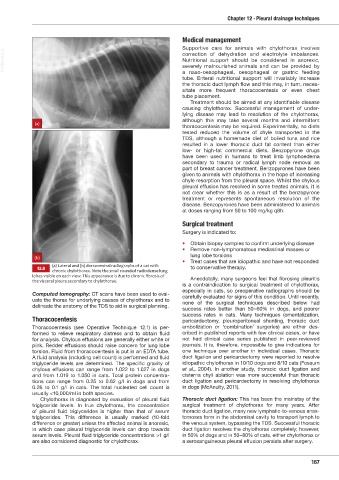
(a) Lateral and (b) dorsoventral radiographs of a cat with
12.9 chronic chylothorax. Note the small rounded radiodense lung to conservative therapy.
lobes visible on each vie . This appearance is due to chronic fibrosis of
the visceral pleura secondary to chylothorax. Anecdotally, many surgeons feel that fibrosing pleuritis
is a contraindication to surgical treatment of chylothorax,
especially in cats, so preoperative radiographs should be
Computed tomography: CT scans have been used to eval- carefully evaluated for signs of this condition. Until recently,
uate the thorax for underlying causes of chylothorax and to none of the surgical techniques described below had
delineate the anatomy of the TDS to aid in surgical planning.
success rates better than 50–60% in dogs, and poorer
success rates in cats. Many techniques (omen talization,
Thoracocentesis pericardectomy, pleuroperitoneal shunting, thoracic duct
Thoracocentesis (see Operative Technique 12.1) is per- embolization or ‘combination’ surgeries) are either des-
formed to relieve respiratory distress and to obtain fluid cribed in published reports with few clinical cases, or have
for analysis. Chylous effusions are generally either white or not had clinical case series published in peer-reviewed
pink. Redder effusions should raise concern for lung lobe journals. It is, therefore, impossible to give indications for
torsion. Fluid from thoracocentesis is put in an EDTA tube. one technique over another in individual cases. Thoracic
A fluid analysis (including cell count) is performed and fluid duct ligation and pericardectomy were reported to resolve
triglyceride levels are determined. The specific gravity of idiopathic chylothorax in 10/10 dogs and 8/10 cats (Fossum
chylous effusions can range from 1.022 to 1.027 in dogs et al., 2004). In another study, thoracic duct ligation and
and from 1.019 to 1.050 in cats. Total protein concentra- cisterna chyli ablation was more successful than thoracic
tions can range from 0.25 to 0.62 g/l in dogs and from duct ligation and pericardectomy in resolving chylothorax
0.26 to 0.1 g/l in cats. The total nucleated cell count is in dogs (McAnulty, 2011).
usually <10,000/ml in both species.
Chylothorax is diagnosed by evaluation of pleural fluid Thoracic duct ligation: This has been the mainstay of the
triglyceride levels. In true chylothorax, the concentration surgical treatment of chylothorax for many years. After
of pleural fluid triglycerides is higher than that of serum thoracic duct ligation, many new lymphatic-to-venous anas-
triglycerides. This difference is usually marked (10-fold tomoses form in the abdominal cavity to transport lymph to
difference or greater) unless the affected animal is anor exic, the venous system, bypassing the TDS. Successful thoracic
in which case pleural triglyceride levels can drop towards duct ligation resolves the chylothorax completely; however,
serum levels. Pleural fluid triglyceride concentrations >1 g/l in 50% of dogs and in 50–80% of cats, either chylothorax or
are also considered diagnostic for chylothorax. a serosanguineous pleural effusion persists after surgery.
167
Ch12 HNT.indd 167 31/08/2018 12:07