
Page 186 - BSAVA Manual of Canine and Feline Head, Neck and Thoracic Surgery, 2nd Edition
P. 186
Chapter 13 · Surgery of the intrathoracic trachea and mainstem bronchi
The distal portion of the trachea and the mainstem
bronchi are less supported by peritracheal adventitial
VetBooks.ir extensive pneumomediastinum and pneumothorax. Again,
tissue, and in this region minor lacerations can produce
the majority of these lacerations are self-limiting.
Severe pneumothorax should be controlled with thora-
cocentesis or thoracic drain placement until the tracheal
laceration seals (see Chapter 12). In the rare instances
where pneumomediastinum and pneumothorax are un-
controllable, or persist after 3–4 days of conserv ative
treatment, further investigations (tracheobronch oscopy)
and explor atory thoracotomy, followed by identification,
debridement and surgical closure of the laceration,
are indicated.
(a)
Non-traumatic tracheal and
bronchial conditions
Tracheal and bronchial foreign bodies
Tracheal foreign bodies are seen more commonly in cats,
whereas bronchial foreign bodies are more common in
outdoor/hunting dogs. Extremely large foreign bodies that
become wedged within the intrathoracic trachea can be
inhaled by cats, which is surprising given the sensitivity of
the feline larynx.
Clinical signs
(b) Typically, these animals have a sudden onset of moist
cough, respiratory noise and varying degrees of dyspnoea.
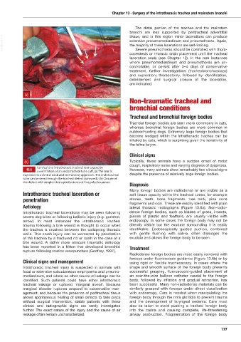
Cervical and intrathoracic tracheal tear caused by
13.5 However, many animals show remarkably few clinical signs
overinflation of an endotracheal tube cuff. a The tear is
exposed via a ventral neck and sternotomy approach. The endotracheal despite the presence of relatively large foreign bodies.
tube can be seen through the tracheal defect (arrowed). (b) Closure of
the defect ith simple interrupted sutures of fine polydio anone.
Diagnosis
Many foreign bodies are radiodense or are visible as a
Intrathoracic tracheal laceration or soft tissue opacity within the tracheal lumen, for example
penetration stones, teeth, bone fragments, tree bark, pine cone
fragments and coal. These are readily identified with plain
Aetiology lateral thoracic radiographs (Figure 13.6a). Non-radio-
Intrathoracic tracheal lacerations may be seen following dense foreign bodies, such as blades of grass, insects,
severe dog bites or following ballistic injury (e.g. gunshot, pieces of plastic and feathers, are usually visible with
arrow). In most instances the intrathoracic tracheal endoscopy. In some cases the foreign body may not be
trauma following a bite wound is thought to occur when directly visible but the exudate surrounding it is easily
the trachea is crushed between the collapsing thoracic identi fiable. Endoscopically guided suction, combined
walls. This crush injury can be worsened by penetration with gentle flushing with saline, often dislodges the
of the trachea by a fractured rib or tooth in the case of a exudate and allows the foreign body to be seen.
bite wound. A rather more obscure traumatic aetiology
has been reported in a kitten that developed bronchial Treatment
rupture following routine venepuncture (Godfrey, 1997).
Radiodense foreign bodies are most easily removed with
forceps under fluoroscopic guidance (Figure 13.6b) or by
Clinical signs and management using rigid or flexible tracheoscopy. In cases where the
Intrathoracic tracheal injury is suspected in animals with shape and smooth surface of the foreign body prevents
focal or extensive subcutaneous emphysema and pneumo- successful grasping, fluoroscopic-guided placement of
mediastinum, and where no other source of leakage can be an over-the-wire balloon catheter caudal to the foreign
identified. Such patients could have either intrathoracic body, followed by inflation and gradual retraction, has
tracheal leakage or ruptured ‘marginal alveoli’. Because been successful. Many non-radiodense materials can be
marginal alveolar ruptures respond to conservative man- similarly grasped with forceps under direct visualization
agement, and because the presence of peritracheal tissue with endoscopy. Care is needed when manipulating the
allows spontaneous healing of small defects to take place foreign body through the rima glottidis to prevent trauma
without surgical intervention, stable patients with these and the development of laryngeal oedema. Care must
clinical and radiographic signs are rarely investigated also be taken to avoid pushing a tracheal foreign body
further. The exact nature of the injury and the cause of air into the carina and causing complete, life-threatening
leakage often remain uncharacterized. airway obstruction. Fragmentation of the foreign body
177
Ch13 HNT.indd 177 31/08/2018 13:01