
Page 314 - Canine Lameness
P. 314
286 18 Tarsal Region
(A) (C) (E) (G)
TARSAL REGION (B) (D) (F)
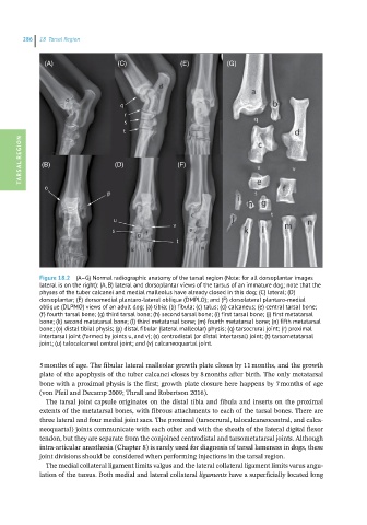
Figure 18.2 (A–G) Normal radiographic anatomy of the tarsal region (Note: for all dorsoplantar images
lateral is on the right): (A, B) lateral and dorsoplantar views of the tarsus of an immature dog; note that the
physes of the tuber calcanei and medial malleolus have already closed in this dog; (C) lateral; (D)
dorsoplantar; (E) dorsomedial plantaro-lateral oblique (DMPLO); and (F) dorsolateral plantaro-medial
oblique (DLPMO) views of an adult dog; (a) tibia; (b) fibula; (c) talus; (d) calcaneus; (e) central tarsal bone;
(f) fourth tarsal bone; (g) third tarsal bone; (h) second tarsal bone; (i) first tarsal bone; (j) first metatarsal
bone; (k) second metatarsal bone; (l) third metatarsal bone; (m) fourth metatarsal bone; (n) fifth metatarsal
bone; (o) distal tibial physis; (p) distal fibular (lateral malleolar) physis; (q) tarsocrural joint; (r) proximal
intertarsal joint (formed by joints u, and v); (s) centrodistal (or distal intertarsal) joint; (t) tarsometatarsal
joint; (u) talocalcaneal central joint; and (v) calcaneoquartal joint.
5 months of age. The fibular lateral malleolar growth plate closes by 11 months, and the growth
plate of the apophysis of the tuber calcanei closes by 8 months after birth. The only metatarsal
bone with a proximal physis is the first; growth plate closure here happens by 7 months of age
(von Pfeil and Decamp 2009; Thrall and Robertson 2016).
The tarsal joint capsule originates on the distal tibia and fibula and inserts on the proximal
extents of the metatarsal bones, with fibrous attachments to each of the tarsal bones. There are
three lateral and four medial joint sacs. The proximal (tarsocrural, talocalcaneocentral, and calca-
neoquartal) joints communicate with each other and with the sheath of the lateral digital flexor
tendon, but they are separate from the conjoined centrodistal and tarsometatarsal joints. Although
intra-articular anesthesia (Chapter 8) is rarely used for diagnosis of tarsal lameness in dogs, these
joint divisions should be considered when performing injections in the tarsal region.
The medial collateral ligament limits valgus and the lateral collateral ligament limits varus angu-
lation of the tarsus. Both medial and lateral collateral ligaments have a superficially located long