
Page 328 - Canine Lameness
P. 328
300 18 Tarsal Region
TARSAL REGION
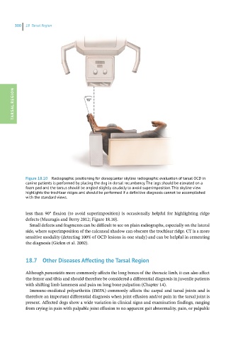
Figure 18.10 Radiographic positioning for dorsoplantar skyline radiographic evaluation of tarsal OCD in
canine patients is performed by placing the dog in dorsal recumbency. The legs should be elevated on a
foam pad and the tarsus should be angled slightly caudally to avoid superimposition. This skyline view
highlights the trochlear ridges and should be performed if a definitive diagnosis cannot be accomplished
with the standard views.
less than 90° flexion (to avoid superimposition) is occasionally helpful for highlighting ridge
defects (Mauragis and Berry 2012; Figure 18.10).
Small defects and fragments can be difficult to see on plain radiographs, especially on the lateral
side, where superimposition of the calcaneal shadow can obscure the trochlear ridge. CT is a more
sensitive modality (detecting 100% of OCD lesions in one study) and can be helpful in cementing
the diagnosis (Gielen et al. 2002).
18.7 Other Diseases Affecting the Tarsal Region
Although panosteitis more commonly affects the long bones of the thoracic limb, it can also affect
the femur and tibia and should therefore be considered a differential diagnosis in juvenile patients
with shifting limb lameness and pain on long bone palpation (Chapter 14).
Immune-mediated polyarthritis (IMPA) commonly affects the carpal and tarsal joints and is
therefore an important differential diagnosis when joint effusion and/or pain in the tarsal joint is
present. Affected dogs show a wide variation in clinical signs and examination findings, ranging
from crying in pain with palpable joint effusion to no apparent gait abnormality, pain, or palpable