
Page 108 - Clinical Small Animal Internal Medicine
P. 108
76 Section 2 Endocrine Disease
Fourteen thyroid glands from euthyroid cats without his released from “pop‐top” canned cat food lids, and con
VetBooks.ir tologically detectable thyroid lesions were examined sumption of commercial canned food. One theory as to
why the number and percentage of unexpected outliers
similarly as controls. Results from these investigations
showed that all cases of nodular follicular hyperplasia/
take several years of exposure to such environmental,
adenomas stained positively for overexpression of c‐Ras becomes accelerated over the age of 9 years is that it may
protein using a mouse monoclonal antihuman pan‐Ras dietary, and genetic factors before they express them
antibody. The most intensely positively staining regions selves clinically and hyperthyroidism ensues, although
were in luminal cells surrounding abortive follicles. this topic requires further investigation.
Subjacent thyroid and parathyroid glands from euthy
roid cats did not stain immunohistochemically for pan‐
Ras. There was no detectable staining for either Bc12 or History and Clinical Signs
p53 in any of the cats. These results indicated that over
expression of c‐ras was highly associated with areas of With time, we have seen both an increase in the diagno
nodular follicular hyperplasia/adenomas of feline thy sis of hyperthyroidism and a decrease in the severity of
roid glands, and mutations in this oncogene may play a the clinical signs associated with thyrotoxicosis. This is
role in the etiopathogenesis of hyperthyroidism in cats. most likely due to an increased awareness on the part of
As with the study on G protein abnormalities, c‐ras the pet owner and the veterinarian as well as the
mutations could either be an initiating cause of hyper increased use of T4 concentrations as an integral part of
thyroidism or simply mediate the effects of an as yet uni routine feline health screening. We have also seen addi
dentified dietary or environmental initiator. tional work on some of the less obvious manifestations of
Alterations in the thyrotropin (TSH) receptor were hyperthyroidism such as hypertension which may be
also examined in cats with hyperthyroidism. The authors clinically silent and/or present initially with ocular signs,
used the polymerase chain reaction (PCR) to amplify as well as the effects of hyperthyroidism on the cardio
codons 480–640 of the previously uncharacterized feline vascular and renal system (to be discussed later).
thyrotropin receptor (TSHR) gene, and determined the As stated earlier, the clinical signs associated with
DNA sequence in this transmembrane domain region. hyperthyroidism have been decreasing in severity over
They then analyzed single‐stranded conformational pol the years (Box 10.1). A paper examined the electrocar
ymorphisms in thyroid DNA from 11 sporadic cases of diographic and radiographic changes seen in hyperthy
feline thyrotoxicosis and leukocyte DNA from two cases roid cats today versus those seen 10–12 years ago. Two
of familial feline thyrotoxicosis. They also determined populations (1992–1993 and 1979–1982) of confirmed
the DNA sequence of this region of the TSHR in five of hyperthyroid cats were compared to determine whether
the cases of sporadic feline thyrotoxicosis and the two the incidence of certain cardiovascular specific manifes
familial thyrotoxic cats. The normal feline TSHR tations of feline thyrotoxicosis had experienced similar
sequence between codons 480 and 640 is highly homolo changes. Sinus tachycardia, which is the most commonly
gous to that of other mammalian TSHRs, with 95%, 92%, recognized cardiac manifestation of feline thyrotoxico
and 90% amino acid identity between the feline receptor sis, was not as prevalent in the 1993 group when com
and canine, human, and bovine TSHRs, respectively. pared to the 1982 group. This was also true for the
Thyroid gland DNA from 11 cats with sporadic thyro finding of an increased R‐wave amplitude on lead II elec
toxicosis did not have mutations in this region of the trocardiography. Both groups had a similar low inci
TSHR gene. Leukocyte DNA from two littermates with dence of atrial and ventricular dysrhythmias; however,
familial feline thyrotoxicosis did not harbor mutations of the 1993 group had a significantly higher occurrence of
this region of the TSHR gene. These studies suggested right bundle branch block. Thoracic radiographs were
that TSHR gene mutations are likely not involved in deemed necessary in a larger proportion of the 1982
feline hyperthyroidism. group compared to the 1993 group. Although there were
Since its first description in 1979, the incidence of
hyperthyroidism has dramatically increased, prompting
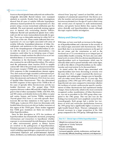
veterinarians and researchers to hypothesize whether Box 10.1 Clinical signs of feline hyperthyroidism
exposure to environmental thyroid‐disruptor chemicals Weight loss and poor hair coat
or other environmental, genetic or dietary factors Aggressive or “cranky” behavior
are involved in the pathogenesis of hyperthyroidism. Periodic vomiting
Potential exposure to several substances has been impli Polyuria and polydipsia
cated, including organohalogen compounds such as pol Increased appetite, activity, restlessness, and heart rate
ychlorinated biphenyls and polybrominated diphenyl Occasionally, difficulty breathing, weakness, and depression
ethers, fertilizers, soy isoflavones, bisphenol‐A primarily