Page 472 - Clinical Small Animal Internal Medicine
P. 472
440  Section 5  Critical Care Medicine

            exposure to EG since many formulations contain a fluo-  ings at different time points following EG ingestion again
  VetBooks.ir  rescent dye. Unfortunately, since some formulations do   demonstrate how important it is to establish a timeline
                                                              of possible exposure.
            not contain added dye, Woods’ lamp evaluation cannot
                                                                The treatment of EG toxicity is similarly dependent on
            be used as a sole diagnostic test. Diagnosis of EG toxicity
            must therefore be made based on clinical suspicion and   timing of exposure. Definitive treatment of EG ingestion
            the presence of certain findings on any of the following   revolves around preventing accumulation of toxic metab-
            tests: osmole gap, anion gap, urinalysis (calcium oxalate   olites. Metabolism of EG to glycolic acid by the enzyme
            crystalluria), blood chemistry, or abdominal ultra-  alcohol dehydrogenase (ADH) is the most important step
            sonography. Ethylene glycol is an effective osmole, and   in the development of EG toxicity. Veterinary patients
            the presence of significant ethylene glycol levels in the   have traditionally been treated with IV ethanol adminis-
            blood can result in neurologic symptoms (e.g., ataxia   tration. Ethanol is metabolized by ADH, and ADH has a
            and stupor) due to hyperosmolality.               higher affinity for ethanol than EG. Administration of
             The presence of significant amounts of EG in the   ethanol, therefore, can limit glycolic acid accumulation.
            blood leads to an increase in the “osmole gap,” which is   At the same time, high rates of IV fluids can be given to
            the difference between measured blood osmolality and   drive urinary excretion of unmetabolized EG molecules.
            calculated blood osmolality (equation 1).         Ethanol administration can be an effective treatment for

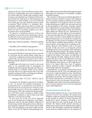
            Osmole gap  Measured osmolalityCalculated osmolality    EG ingestion, but long courses of treatment are needed
                                                              and side‐effects (including hyperosmolality, CNS signs
                                                     (Eq. 1)  such as sedation, and GI signs such as vomiting) are fre-
              Osmolality can be estimated using equation 2.   quently observed. ADH inhibitors such as 4‐methyl-
                                                              pyrazole (4‐MP, also known as fomepazole) prevent
             186   Na K     BUN/.      Glu/ 18 /.             metabolism of EG to toxic metabolites while offering less
              .
                                  2 8
                                                0 93 (Eq. 2)
                                                              risk of side‐effects than ethanol. The primary drawbacks
            The normal whole‐blood osmole gap has been reported   to 4‐MP are limited availability and relatively high cost
            as 3.49 mOsm and the normal plasma osmole gap as   compared with ethanol. Cats require very high doses of
            –2.01 mOsm. A significant increase in the osmole gap in   4‐MP compared with dogs, making cats more likely to
            a patient who may have been exposed to EG or who is   develop side‐effects such as CNS depression and ataxia
            showing symptoms should raise clinical suspicion for EG   following treatment with 4‐MP. Treatment of cats with
            poisoning.                                        ADH inhibition using ethanol or 4‐MP is typically inef-
             Fatalities due to EG toxicity are caused by acute renal   fective  if not  begun  within three  hours of  ingestion,
            failure as EG metabolism leads to accumulation of   whereas treatment of dogs is typically successfully if
              metabolic acids such as glycolic and oxalic acid. This accu-  started within eight hours of ingestion.
            mulation leads to a metabolic acidosis and an   elevated   Hemodialysis is a very effective treatment for removal
            anion gap (AG) seen at approximately three hours after   of EG and glycolic acid; 90–95% of circulating EG can be
            ingestion, peaking at six hours, and lasting up to 48 hours.   removed in a single hemodialysis session. Intermittent
            AG can be determined using equation 3.            hemodialysis may also effectively treat azotemia and
                                                              hyperkalemia in patients with acute kidney injury from
                 Anion gap  Na    K     Cl  HCO3   (Eq. 3)    EG toxicity, allowing time to determine if recovery of

                                                              kidney function occurs over the span of several weeks.
             Normal AG is 12–24 mEq/L in dogs and 13–27 mEq/L   Regardless of the mode of treatment, prompt interven-
            in cats. An elevated AG indicates accumulation of   tion is of utmost importance due to the time‐dependent
            unmeasured anions. An elevated AG is not specific to   nature of this lethal toxin.
            EG intoxication, but an elevated AG 3–6 hours or more
            after possible exposure to EG may be considered a sign   Uncertain Exposure to a Known Toxin
            of EG exposure. The oxalic acid formed during metabo-
            lism of EG binds calcium, resulting in calcium oxalate   When treating a patient who may or may not have been
            monohydrate crystalluria as early as three hours after   exposed to a known toxic substance, the clinician must
            ingestion in cats (six hours in dogs).            formulate a diagnostic testing and treatment plan based
             In some cases, EG toxicity may not be diagnosed   on the presence or absence of clinical symptoms, the
            prior  to  the  development  of  oligoanuric  renal  failure.   likelihood of clinical symptoms developing if toxic
            Progressive azotemia may help establish a diagnosis of   exposure did in fact occur, and the danger of the toxin
            EG poisoning, but this finding almost always indicates a   involved. The clinician should consider contacting an
            grave prognosis. Diagnosis of EG toxicity must be made   animal poison control center to obtain the most extensive
            prior to the development of azotemia to allow the best   and up‐to‐date information regarding the probability of
            chance at survival. The different clinicopathologic find-  toxicity from any possible exposure to the substance in

