
Page 598 - Clinical Small Animal Internal Medicine
P. 598
566 Section 6 Gastrointestinal Disease
(a)
VetBooks.ir Upper esophageal sphincter
Swallow
Relaxation
Thorax
Esophagus
Peristaltic wave
Gastric Lower esophageal sphincter
(b)
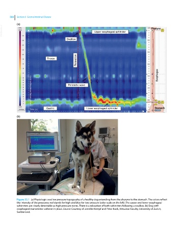
Figure 53.1 (a) Physiologic swallow pressure topography of a healthy dog extending from the pharynx to the stomach. The colors reflect
the intensity of the pressures; red stands for high and blue for low pressure (color scale on the left). The upper and lower esophageal
sphincters are clearly detectable as high‐pressure zones. There is a relaxation of both sphincters following a swallow. (b) Dog with
esophageal manometer catheter in place. Source: Courtesy of Jennifer Kempf and Peter Kook, Vetsuisse Faculty, University of Zurich,
Switzerland.