
Page 817 - Withrow and MacEwen's Small Animal Clinical Oncology, 6th Edition
P. 817
CHAPTER 34 Miscellaneous Tumors 795
Breed differences have also been studied. Two studies have
found that FCR are more likely to have HS of the limbs and
Localized HS is
530,535
BMD have more diffuse visceral disease.
VetBooks.ir seven times more frequent in the FCR than in the BMD and dis-
seminated HS is two times more frequent in the BMD than the
FCR. FCR with HS were also older at diagnosis than BMDs with
HS in one study (mean 8.6 vs 7.7 years). 530 Differences in histo-
pathology between these breeds have also been described. 536
HS may present with either localized or loco-regional organ
involvement or with a disseminated/multiorgan involvement. HS
is the preferred term identifying malignant tumors of histiocytic
origin, and the older term “malignant histiocytosis” refers specifi-
cally to the disseminated form of the disease. Reported anatomic
sites include lung, LN, liver, spleen, stomach, pancreas, medias-
tinum, skin, skeletal muscle, central nervous system, bone, bone
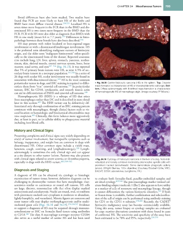
marrow, nasal cavity, and eyes 537–546 (Fig. 34.13). It was reported
that 5% of primary brain tumors were HS, as were 4.5% of sec-
ondary brain tumors in a necropsy population. 547,548 In a series of
26 dogs with ocular HS, ocular involvement was usually found in
association with disseminated disease. 543 HS is the most common
periarticular tumor in dogs, and IHC staining is required to differ- • Fig. 34.13 Canine histiocytic sarcoma (HS) in the spleen. Top. Discrete
entiate them from “true” synovial cell sarcoma. 538 In periarticular mass formation is characteristic of HS of interstitial dendritic cell origin. Bot-
tumors, IHC for CD18, cytokeratin, and smooth muscle actin tom. Diffuse splenomegaly with ill-defined mass formation is characteristic
can aid in differentiation of PAHS and synovial cell sarcoma. 538 of hemophagocytic HS of macrophage origin. (Image courtesy PF Moore.)
Hemophagocytic HS (HHS) is a subtype of HS that arises
from macrophages rather than DC and is described in more detail
later in this section. 483 The HHS variant can be definitively dif-
ferentiated only through confirmation of an IHC staining pattern
consistent with macrophages, though clinical factors such as vis-
ceral location or hematologic and biochemical abnormalities may
raise suspicion. 483 Clinically, this form behaves more aggressively
due, at least in part, to its cellular ability to phagocytose material
including host blood cells.
History and Clinical Signs
Presenting complaints and clinical signs vary widely depending on
site(s) of tumor involvement, but nonspecific symptoms such as
lethargy, inappetence, and weight loss are common in dogs with
disseminated HS. Other common signs include a visible mass,
lameness, cough, vomiting, and lymphadenomegaly. 528 Lymph-
adenomegaly is sometimes the only clinical sign and can appear
at a site distant to other tumor lesions. Patients may also present 20 m
with clinical signs related to severe anemia or thrombocytopenia, • Fig. 34.14 Cytology of histiocytic sarcoma in the liver of a dog. Note indi-
especially in dogs with the HHS variant. 483,540–542 vidualized and loosely cohesive moderately pleomorphic spindle cells with
prominent nucleoli (arrowheads). Some demonstrate phagocytic activity
Diagnosis and Staging (arrow). (Wright-Giemsa, 100× objective.) (Courtesy Elizabeth Little, VMD,
DACVP, IDEXX Laboratories, Langhorne, PA.)
A diagnosis of HS can be obtained via cytologic or histologic
examination of tumor tissue; however, definitive diagnosis can be to evaluate both formalin-fixed, paraffin-embedded samples and
challenging in pleomorphic tumors that have morphologic char- air-dried cytology. 499,548 The pan-macrophage marker ionized cal-
acteristics similar to carcinomas or round cell tumors. HS cells cium-binding adapter molecule 1 (Iba1) also appears to have utility
are large, discrete, mononuclear cells that often display marked as a marker of cells of monocyte and macrophage lineage, though
anisocytosis and anisokaryosis. Nuclei are round, oval, or reniform it cannot differentiate the various histiocytic disorders. 549 If fresh
with prominent nucleoli and cytoplasm is moderate to abundant, or frozen tissue is available, further confirmation and subclassifica-
lightly basophilic, and vacuolated. Mitotic figures are common and tion of the cell of origin can be performed using antibody staining
f
some tumor cells may display erythrophagocytosis and/or multi- for CD1 or the CD11 α subunits. 478,480 Recently, the CADET
nucleated giant cells (Figs. 34.14 and 34.15). 478,480,547 Evidence histiocytic malignancy assay has become commercially available.
to support a diagnosis of HS may be acquired through immuno- Using this assay, tumor biopsy or cytology samples are evaluated
cytochemistry or IHC on formalin fixed tissues using antibodies for copy number aberrations consistent with those found in cases
to CD18. 480 The class A macrophage scavenger receptor CD204 of confirmed HS. The sensitivity and specificity of the assay have
also serves as a useful marker of canine HS and has been used been estimated to be 78% and 95%, respectively. 530,550