
Page 214 - BSAVA Manual of Canine and Feline Head, Neck and Thoracic Surgery, 2nd Edition
P. 214
Chapter 16 · Surgery of the mediastinum
Pneumomediastinum Mediastinal neoplasia
VetBooks.ir Conditions causing pneumomediastinum that require Soft tissue masses within the mediastinum are rare, but
when present they can usually be seen readily on plain
surgery are dealt with elsewhere in this book (see Chapters
thoracic radiographs (see Figures 16.5, 16.6 and 16.8).
9, 13 and 14). Pneumomediastinum can occur as a sponta-
neous event secondary to pulmonary pathology, severe
majority of masses observed.
dyspnoea and/or coughing, or following rupture of the Primary and metastatic neoplastic lesions account for the
oesophagus, trachea, mainstem bronchi or marginal
alveoli. Road traffic accidents or bite wounds are the
common traumatic insults, whereas mechanical ventilation,
transtracheal aspiration, tracheostomy tube placement and
endotracheal intubation are the typical iatrogenic events
leading to pneumomediastinum (Brown and Holt 1995;
Jordan et al., 2013). Pneumomediastinum may also result
from migration of air within the cervical fascia, as observed
following rupture of the cervical trachea. Pneumothorax,
subcutaneous emphysema and pneumo peritoneum may
all develop as a result of air leakage into the mediastinum.
Whereas pneumothorax may develop secondary to
pneumomediastinum, the converse is extremely unlikely.
Although theoretically a rapidly forming pneumomedia-
stinum could cause pressure on mediastinal vessels,
reducing venous return to the heart, the flimsy nature of the
canine and feline mediastinum means that pneumothorax
develops prior to mediastinal tamponade. Consequently,
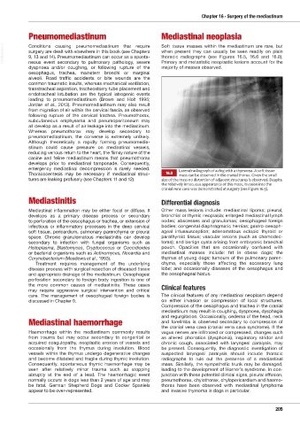
emergency mediastinal decompression is rarely needed. Lateral radiograph of a dog with a thymoma. A soft tissue
Thoracocentesis may be necessary if media stinal struc- 16.8 mass can be observed in the cranial thorax. Given the small
tures are leaking profusely (see Chapters 11 and 12). size of the mass no distortion of adjacent structures is observed. Despite
the relatively innocuous appearance of this mass, invasion into the
cranial vena cava was demonstrated at surgery (see Figure 16.9).
Mediastinitis Differential diagnosis
Mediastinal inflammation may be either focal or diffuse. It Other mass lesions include: mediastinal lipoma; pleural,
develops as a primary disease process or secondary bronchial or thymic neoplasia; enlarged mediastinal lymph
to perforation of the oesophagus or trachea, or extension of nodes; abscesses and granulomas; oesophageal foreign
infectious or inflammatory processes in the deep cer vical bodies; congenital diaphragmatic hernias; gastro-oesoph-
soft tissue, pericardium, pulmonary paren chyma or pleural ageal intussusception; adenomatous ectopic thyroid or
space. Chronic granulomatous media stinitis can develop parathyroid tissue; vascular lesions (such as chemodec-
secondary to infection with fungal organisms such as toma); and benign cysts arising from embryonic branchial
Histoplasma, Blastomyces, Crypto coccus or Coc cidioides pouch. Opacities that are occasionally confused with
or bacterial organisms such as Actino myces, Nocardia and mediastinal masses include: fat in obese dogs; the
Corynebacterium (Meadows et al., 1993). thymus of young dogs; tumours of the pulmonary paren-
Treatment requires management of the underlying chyma, especially those affecting the accessory lung
disease process with surgical resection of diseased tissue lobe; and occasionally diseases of the oesophagus and
and appropriate drainage of the mediastinum. Oesophageal the oesophageal hiatus.
perforation secondary to foreign body ingestion is one of
the more common causes of mediastinitis. These cases Clinical features
may require aggressive surgical intervention and critical
care. The management of oesophageal foreign bodies is The clinical features of any mediastinal neoplasm depend
discussed in Chapter 9. on either invasion or compression of local structures.
Compression of the oesophagus and trachea in the cranial
mediastinum may result in coughing, dyspnoea, dysphagia
and regurgitation. Occasionally, oedema of the head, neck
Mediastinal haemorrhage and forelimbs is observed secondary to compression of
the cranial vena cava (cranial vena cava syndrome). If the
Haemorrhage within the mediastinum commonly results vagus nerves are infiltrated or compressed, changes such
from trauma but may occur secondary to congenital or as altered phonation (dysphonia), inspiratory stridor and
acquired coagulopathy, neoplastic erosion of vessels and chronic cough, associated with laryngeal paralysis, may
occasionally from the thymus during involution. Blood be present. Consequently, the diagnostic investigation of
vessels within the thymus undergo degenerative changes suspected laryngeal paralysis should include thoracic
and become dilatated and fragile during thymic involution. radiographs to rule out the presence of a mediastinal
Consequently, spontaneous thymic haemorrhage may be mass. Similarly, the sympathetic trunk may be damaged,
seen after relatively minor trauma such as stopping leading to the development of Horner’s syndrome. In con-
abruptly at the end of a lead. The haemorrhagic event junction with these potential clinical signs, pleural effusion,
normally occurs in dogs less than 2 years of age and may pneumothorax, chylothorax, chylopericardium and haemo-
be fatal. German Shepherd Dogs and Cocker Spaniels thorax have been observed with mediastinal lymphoma
appear to be over-represented. and invasive thymoma in dogs in particular.
205
Ch16 HNT.indd 205 31/08/2018 13:32