
Page 222 - BSAVA Manual of Canine and Feline Head, Neck and Thoracic Surgery, 2nd Edition
P. 222
Chapter 17 · Surgery of the diaphragm
the diaphragm. If there is tension on the closure, the
ventral border of the defect may be approximated to
VetBooks.ir mattress sutures.
the abdominal fascia over the costal arch with horizontal
In the majority of animals, there is sufficient tissue
to allow closure of the defect without undue tension. In
animals with a large defect, the pericardial sac is incised
cranial to the junction of the diaphragm and pericardium,
and this extra ring of tissue is used to close the defect.
Alternatively, the diaphragm may be incised on each side
of the defect at its paracostal attachment and bilateral
rotation flaps used to close the defect (Bellah et al.,
1989). Umbilical hernias and defects in the cranioventral
abdominal wall are closed during closure of the lapar-
otomy wound.
Postoperative care and complications: Postoperative
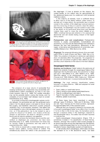
Peritoneopericardial diaphragmatic hernia: lateral view of the recovery is usually uncomplicated. Complications are
17.4 thorax following oral administration of barium suspension to
the dog in Figure 17.3. Barium-filled loops of small intestine are present uncommon, but may include haemorrhage from adhesions
within the pericardium and outline a cranial ventral abdominal hernia. between the liver and pericardium, dehiscence of the
repair, re-herniation and development of constrictive peri-
carditis (Wallace et al., 1992; Burns et al., 2013).
Prognosis: The prognosis following closure of an uncompli-
cated PPDH is good (Evans and Biery, 1980; Hay et al.,
1989). The presence of sternal and abdominal wall defects
does not adversely affect the prognosis. The prognosis for
animals with concurrent congenital heart defects is poorer
and the outlook depends on the nature of the heart disease.
Oesophageal hiatal hernia
Anatomy and incidence: Hiatal hernia is the protrusion of
abdominal contents through the oesophageal hiatus of the
diaphragm into the thoracic cavity. It is uncommon in dogs
and rare in cats (Ellison et al., 1987; Waldron et al., 1990).
Shar-Peis appear to be predisposed to this condition
(Prymak et al., 1989; Williams, 1990; Callan et al., 1993) and
it is most commonly seen in young brachycephalic dogs.
Peritoneopericardial diaphragmatic hernia: visualization of
17.5 the heart through the defect in the diaphragm. Aetiology: The most common classification system
favoured in humans (Skinner, 1986) and adopted for small
animals is as follows:
The presence of a large volume of pericardial fluid
causing cardiovascular compromise may necessitate peri- • Type I: sliding or axial hiatal hernia
cardiocentesis before anaesthesia, but this is not com- • Type II: rolling or para-oesophageal hiatal hernia
monly required (Hay et al., 1989). Full cardiac evaluation • Type III: combined type I and type II
may not be possible before closing the hernia, and the • Type IV: herniation of other organs into the thorax, e.g.
possibility of concomitant heart disease must be consid- intestine, spleen or pancreas.
ered (Feldman et al., 1968).
In contrast to a traumatic diaphragmatic rupture, contin - All these types have been described in animals,
uity between the pericardial sac and the peritoneal cavity although types III (Williams, 1990) and IV (Brinkley, 1990)
means that the pleural space is not open to the air during are rare. Type I hernias are the most common, represent-
the surgery, and therefore intermittent positive pressure ing approximately 90% of the reported cases.
ventilation may not be required. However, if the defect has Although they are frequently referred to synonymously,
to be enlarged to return the herniated viscera to the abdo- a true para-oesophageal hiatal hernia differs from the type
men, or if the pericardium has to be incised to use a II hernia in that, in the former case, the herniation occurs
portion for closure of the defect, then incision of the dia- through a separate defect in the diaphragm, adjacent to
phragm or pericardium will result in iatrogenic pneumo- the oesophageal hiatus. This defect has not been reported
thorax and will require ventilatory management. in small animals.
Adhesions between the herniated viscera and the peri- Gastro-oesophageal intussusception (Figure 17.6) is
cardium or diaphragm are uncommon, but have been sometimes included in the classification of hiatal hernias.
described (Hay et al., 1989). Incarcerated liver lobes may However, although this condition does involve passage of
be necrotic, fragile or lipomatous, and may need to be an abdominal organ across the diaphragm into the thoracic
resected (Hay et al., 1989). cavity, it might be argued that it is not a true hernia. It is not
The hernia is closed with a simple interrupted or con- included in human classification systems of hiatal hernia.
tinuous suture pattern, running dorsal to ventral. This An abnormally short oesophagus, which does not allow
simultaneously closes the defect in the pericardium and the stomach to lie in the abdominal cavity, has been
213
Ch17 HNT.indd 213 31/08/2018 13:45