
Page 1203 - Small Animal Clinical Nutrition 5th Edition
P. 1203
Nutrition of Reptiles 1253
Bibliography
VetBooks.ir Boyer TH. Metabolic bone disease. In: Mader DR, ed. Reptile
Medicine and Surgery. Philadelphia, PA: WB Saunders Co,
1996; 385-392.
Mader DR. Use of calcitonin in green iguanas, Iguana iguana,
with metabolic bone disease. Bulletin of the Association of Rep-
tilian and Amphibian Veterinarians 1993; 3: 41.
Rossi J. A practical and effective treatment for metabolic bone
disease in the green iguana. In: Proceedings. North American
Veterinary Conference, Orlando FL, 1992: 707.
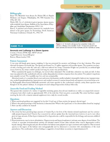
Figure 1. An 18-month-old iguana that presented initially with
CASE 71-3 anorexia, lethargy and a swollen mouth. The iguana’s growth had
been severely stunted and it had markedly swollen mandibular bones.
Anorexia and Lethargy in a Green Iguana
Connie J. Orcutt, DVM, Dipl. ABVP (Avian)
Angell Memorial Animal Hospital
Boston, Massachusetts, USA
Patient Assessment
A two-year-old female green iguana weighing 1.6 kg was presented for anorexia and lethargy of six days’ duration. The owner
obtained the iguana 18 months ago.The iguana was housed in a 75-gallon aquarium with another iguana.The aquarium was heat-
ed with a heating pad under the tank and a 220-watt infrared heat lamp. Ultraviolet-B light was provided by an artificial light
source; however, the owner couldn’t specify how long the source was provided daily.
When examined, the iguana was lethargic, but moved when stimulated. The overall skin coloration was dark and dull. A firm
mass was palpated in the caudodorsal coelomic cavity; deep palpation elicited a response from the patient.The patient’s long bones
were palpably normal. The mandible was firm and non-compressible.
Abnormal results of a complete blood count and serum biochemistry profile included a heterophilic leukocytosis, hyperproteine-
mia,marked hyperphosphatemia,hyperuricemia and marked increases in creatine kinase levels and aspartate aminotransferase activ-
ity. Radiographs demonstrated bilaterally symmetric soft tissue opacities in the caudodorsal coelomic cavity; these opacities were
thought to be the patient’s kidneys. A sonogram revealed enlarged hyperechoic kidneys bilaterally.
Assess the Food and Feeding Method
The iguana’s diet consisted of a variety of vegetables including greens, broccoli and dandelions as well as an unspecified commer-
cial iguana food, which made up approximately 50% of the total diet. Fruit was given occasionally. The owner had been supple-
menting the diet with a vitamin-mineral supplement twice daily for several months.
Questions
1. What nutritional problems are suggested by the diet? Could any of these explain the iguana’s clinical signs?
2. What is the pathophysiology of the biochemical abnormalities? What is the significance of the abnormalities found by imaging?
3. How can this condition be treated?
Answers and Discussion
1. The owner has been providing large amounts of vitamin D in the form of the vitamin-mineral supplement and the commer-
3
cially prepared iguana food. Vitamin D is essential for calcium uptake from the intestinal tract; however, it can be toxic when
3
given in large amounts. Either hypervitaminosis D or hypocalcemia could be responsible for the lethargy and anorexia exhibited
by the patient.
2. Hyperproteinemia may be due to dehydration. Hyperuricemia and hyperphosphatemia indicate some degree of renal failure.The
increase in aspartate aminotransferase activity and elevated creatine kinase value indicate either muscle degeneration and/or
hepatic damage. Renomegaly and pain elicited when the kidneys were palpated are signs consistent with nephrosis.The increased
opacity in the region of the kidneys seen radiographically and by ultrasound is consistent with mineralization of the renal
parenchyma. The combination of these findings is suggestive of renal failure secondary to hypervitaminosis D.