
Page 843 - Small Animal Clinical Nutrition 5th Edition
P. 843
874 Small Animal Clinical Nutrition
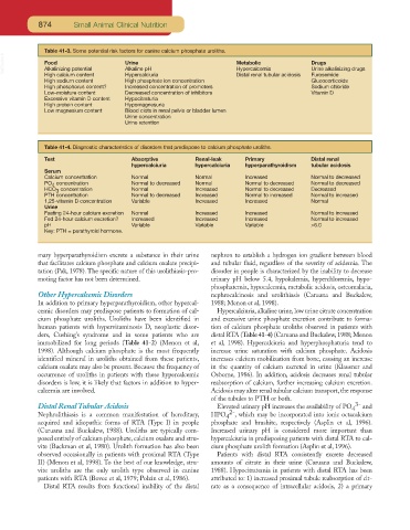
Table 41-3. Some potential risk factors for canine calcium phosphate uroliths.
VetBooks.ir Food Urine Metabolic Drugs
Alkalinizing potential
Urine alkalinizing drugs
Hypercalcemia
Alkaline pH
High calcium content
High sodium content Hypercalciuria Distal renal tubular acidosis Furosemide
Glucocorticoids
High phosphate ion concentration
High phosphorus content? Increased concentration of promoters Sodium chloride
Low-moisture content Decreased concentration of inhibitors Vitamin D
Excessive vitamin D content Hypocitraturia
High protein content Hypomagnesuria
Low magnesium content Blood clots in renal pelvis or bladder lumen
Urine concentration
Urine retention
Table 41-4. Diagnostic characteristics of disorders that predispose to calcium phosphate uroliths.
Test Absorptive Renal-leak Primary Distal renal
hypercalciuria hypercalciuria hyperparathyroidism tubular acidosis
Serum
Calcium concentration Normal Normal Increased Normal to decreased
PO concentration Normal to decreased Normal Normal to decreased Normal to decreased
4
HCO concentration Normal Increased Normal to decreased Decreased
3
PTH concentration Normal to decreased Increased Normal to increased Normal to increased
1,25-vitamin D concentration Variable Increased Increased Normal
Urine
Fasting 24-hour calcium excretion Normal Increased Increased Normal to increased
Fed 24-hour calcium excretion? Increased Increased Increased Normal to increased
pH Variable Variable Variable >6.0
Key: PTH = parathyroid hormone.
mary hyperparathyroidism excrete a substance in their urine nephron to establish a hydrogen ion gradient between blood
that facilitates calcium phosphate and calcium oxalate precipi- and tubular fluid, regardless of the severity of acidemia. The
tation (Pak, 1978). The specific nature of this urolithiasis-pro- disorder in people is characterized by the inability to decrease
moting factor has not been determined. urinary pH below 5.4, hypokalemia, hyperchloremia, hypo-
phosphatemia, hypocalcemia, metabolic acidosis, osteomalacia,
Other Hypercalcemic Disorders nephrocalcinosis and urolithiasis (Caruana and Buckalew,
In addition to primary hyperparathyroidism, other hypercal- 1988; Menon et al, 1998).
cemic disorders may predispose patients to formation of cal- Hypercalciuria, alkaline urine,low urine citrate concentration
cium phosphate uroliths. Uroliths have been identified in and excessive urine phosphate excretion contribute to forma-
human patients with hypervitaminosis D, neoplastic disor- tion of calcium phosphate uroliths observed in patients with
ders, Cushing’s syndrome and in some patients who are distal RTA (Table 41-4) (Caruana and Buckalew,1988; Menon
immobilized for long periods (Table 41-2) (Menon et al, et al, 1998). Hypercalciuria and hyperphosphaturia tend to
1998). Although calcium phosphate is the most frequently increase urine saturation with calcium phosphate. Acidosis
identified mineral in uroliths obtained from these patients, increases calcium mobilization from bone, causing an increase
calcium oxalate may also be present. Because the frequency of in the quantity of calcium excreted in urine (Klausner and
occurrence of uroliths in patients with these hypercalcemic Osborne, 1986). In addition, acidosis decreases renal tubular
disorders is low, it is likely that factors in addition to hyper- reabsorption of calcium, further increasing calcium excretion.
calcemia are involved. Acidosis may alter renal tubular calcium transport, the response
of the tubules to PTH or both.
Distal Renal Tubular Acidosis Elevated urinary pH increases the availability of PO 4 3- and
Nephrolithiasis is a common manifestation of hereditary, HPO 4 2- , which may be incorporated into ionic octacalcium
acquired and idiopathic forms of RTA (Type I) in people phosphate and brushite, respectively (Asplin et al, 1996).
(Caruana and Buckalew, 1988). Uroliths are typically com- Increased urinary pH is considered more important than
posed entirely of calcium phosphate, calcium oxalate and stru- hypercalciuria in predisposing patients with distal RTA to cal-
vite (Backman et al, 1980). Urolith formation has also been cium phosphate urolith formation (Asplin et al, 1996).
observed occasionally in patients with proximal RTA (Type Patients with distal RTA consistently excrete decreased
II) (Menon et al, 1998). To the best of our knowledge, stru- amounts of citrate in their urine (Caruana and Buckalew,
vite uroliths are the only urolith type observed in canine 1988). Hypocitratemia in patients with distal RTA has been
patients with RTA (Bovee et al, 1979; Polzin et al, 1986). attributed to: 1) increased proximal tubule reabsorption of cit-
Distal RTA results from functional inability of the distal rate as a consequence of intracellular acidosis, 2) a primary