
Page 907 - Small Animal Clinical Nutrition 5th Edition
P. 907
Feline Lower Urinary Tract Diseases 939
effects of stress, patients with FIC had significantly increased
VetBooks.ir plasma concentrations of catecholamines, including norepi-
nephrine, compared with healthy cats (Westropp et al, 2006a).
Abnormalities also have been identified in the hypothalam-
ic-pituitary-adrenal axis of cats with FIC.Those with FIC had
significantly decreased cortisol response to administration of
synthetic adrenocorticotropic hormone compared with that of
healthy cats (Westropp et al, 2003). In the same study, adrenal
gland volume was significantly lower in cats with FIC com-
pared with healthy cats; however, there were no correlations
between adrenal size and cortisol production (Westropp et al,
2003, 2005). In another study of the effects of stress, there was
no significant difference in urinary cortisol:creatinine ratios
between patients with FIC and healthy cats (Westropp et al,
2006a). Based on studies conducted to date, it appears there is
dissociation between the response of the sympathetic nervous
system and the hypothalamic-pituitary-adrenal axis to stress in
patients with FIC.
Although many risk factors have been identified in patients
with FIC, additional evaluation is likely needed to identify its
definitive cause(s). Based on current understanding of patho-
genesis, it appears that abnormalities are not limited to the uri-
nary bladder and interactions between other systems (e.g.,
nervous and endocrine) are likely involved. This possibility
must be considered when formulating a treatment plan, which
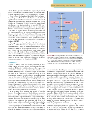
should include a multimodal approach (i.e.,decreasing environ- Figure 46-9. Increasing saturation of urine with urolith-forming con-
mental stress, nutritional management, behavioral modifica- stituents (e.g., calcium, oxalate) results in crystal growth, aggrega-
tion, pain management) for all patients with FIC. tion and ultimately urolith formation, if the components are retained
in the urinary tract. (Adapted from Bartges JW, Osborne CA, Lulich
JP, et al. Methods for evaluating treatment of uroliths. Veterinary
Urolithiasis Clinics of North America: Small Animal Practice 1999; 29: 46.)
Uroliths, or urinary calculi, are composed primarily of crys-
talline mineral concretions with a small amount of organic
matrix. Urolithiasis is a multifaceted process that begins with
microcrystals in urine and ends with mature uroliths some- are not included in these diagrams because they differ for each
where in the urinary tract (Osborne and Kruger, 1984). Urolith of the urolith components and measurement technique; how-
formation occurs in two separate phases-initiation of the crys- ever, the general features apply to all crystalline materials. At
tal nidus and continued growth to form a urolith. In general, concentrations below the solubility product (i.e., in the under-
there are four factors involved in formation of uroliths: 1) over- saturation zone), it is impossible for crystals to form and crys-
saturation of urine with calculogenic crystalloids, which may tals added to such a solution would dissolve. However, crystals
result from increased urinary excretion of these substances or may grow if added to a solution with a concentration greater
increased urine concentration (e.g., due to decreased water than the solubility product. The formation product is the con-
intake), 2) decreased solubility of crystalloids in urine (e.g., centration at which crystals will begin to spontaneously precip-
struvite is less soluble in alkaline urine), 3) presence or absence itate in the absence of preformed crystalline material. Although
of crystallization inhibiters or promoters in urine and 4) reten- the solubility product is constant for a pure crystalline materi-
tion of crystals/uroliths within the urinary tract. al, the formation product is much more difficult to demon-
In its simplest form, initiation and growth of uroliths strate; therefore, this area is illustrated by a shaded band rather
involves chemical precipitation of dissolved ions or molecules than a line (Figure 46-11). Strictly speaking, the ionic activities
from a solution that has become oversaturated with respect to (not concentrations) of the species govern the described solu-
those components (Figure 46-9). From a physiochemical per- bility principles. Ionic activities are influenced by the presence
spective, the degree of saturation or undersaturation of urine of other ions in solution (i.e., ionic strength) and by the pres-
influences the probability that precipitates will form, or if ence of other species that form complex ions, thereby reducing
already present, will dissolve. Relatively simple diagrams depict their “free” concentrations in solution.
states of saturation of any solution (Figures 46-10 and 46-11). The metastable zone is of most interest clinically; in this con-
These diagrams provide the framework for understanding the centration range crystal growth and aggregation may occur or
concept of how nutritional management influences probability other factors (e.g., crystallization inhibitors) may impede or
of urolith formation or dissolution. Units and numerical values prevent crystallization (Figure 46-11). Risk factor reduction