
Page 551 - Feline diagnostic imaging
P. 551
564 31 Body Wall
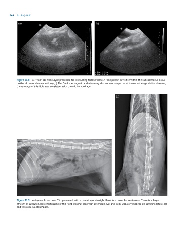
(a) (b)
Figure 31.8 A 7-year-old Himalayan presented for a recurring fibrosarcoma. A fluid pocket is visible within the subcutaneous tissue
on the ultrasound examination (a,b). The fluid is echogenic and a forming abscess was suspected at the recent surgical site. However,
the cytology of this fluid was consistent with chronic hemorrhage.
(b)
(a)
Figure 31.9 A 4-year-old outdoor DSH presented with a recent injury to right flank from an unknown trauma. There is a large
amount of subcutaneous emphysema of the right inguinal area with extension over the body wall as visualized on both the lateral (a)
and ventrodorsal (b) images.