
Page 571 - Feline diagnostic imaging
P. 571
32.1 uvenile one Disease 585
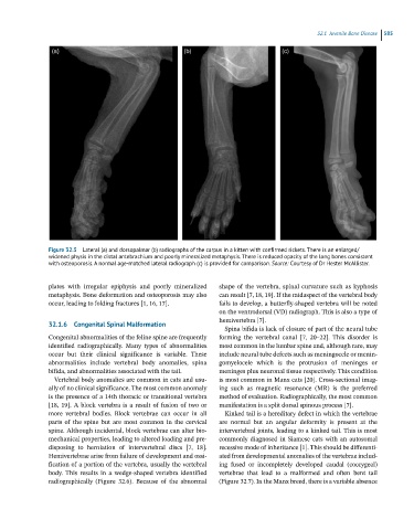
Figure 32.5 Lateral (a) and dorsopalmar (b) radiographs of the carpus in a kitten with confirmed rickets. There is an enlarged/
widened physis in the distal antebrachium and poorly mineralized metaphysis. There is reduced opacity of the long bones consistent
with osteoporosis. A normal age-matched lateral radiograph (c) is provided for comparison. Source: Courtesy of Dr Hester McAllister.
plates with irregular epiphysis and poorly mineralized shape of the vertebra, spinal curvature such as kyphosis
metaphysis. Bone deformation and osteoporosis may also can result [7, 18, 19]. If the midaspect of the vertebral body
occur, leading to folding fractures [1, 16, 17]. fails to develop, a butterfly-shaped vertebra will be noted
on the ventrodorsal (VD) radiograph. This is also a type of
hemivertebra [7].
32.1.6 Congenital Spinal Malformation
Spina bifida is lack of closure of part of the neural tube
Congenital abnormalities of the feline spine are frequently forming the vertebral canal [7, 20–22]. This disorder is
identified radiographically. Many types of abnormalities most common in the lumbar spine and, although rare, may
occur but their clinical significance is variable. These include neural tube defects such as meningocele or menin-
abnormalities include vertebral body anomalies, spina gomyelocele which is the protrusion of meninges or
bifida, and abnormalities associated with the tail. meninges plus neuronal tissue respectively. This condition
Vertebral body anomalies are common in cats and usu- is most common in Manx cats [20]. Cross-sectional imag-
ally of no clinical significance. The most common anomaly ing such as magnetic resonance (MR) is the preferred
is the presence of a 14th thoracic or transitional vertebra method of evaluation. Radiographically, the most common
[18, 19]. A block vertebra is a result of fusion of two or manifestation is a split dorsal spinous process [7].
more vertebral bodies. Block vertebrae can occur in all Kinked tail is a hereditary defect in which the vertebrae
parts of the spine but are most common in the cervical are normal but an angular deformity is present at the
spine. Although incidental, block vertebrae can alter bio- intervertebral joints, leading to a kinked tail. This is most
mechanical properties, leading to altered loading and pre- commonly diagnosed in Siamese cats with an autosomal
disposing to herniation of intervertebral discs [7, 18]. recessive mode of inheritance [1]. This should be differenti-
Hemivertebrae arise from failure of development and ossi- ated from developmental anomalies of the vertebrae includ-
fication of a portion of the vertebra, usually the vertebral ing fused or incompletely developed caudal (coccygeal)
body. This results in a wedge-shaped vertebra identified vertebrae that lead to a malformed and often bent tail
radiographically (Figure 32.6). Because of the abnormal (Figure 32.7). In the Manx breed, there is a variable absence