
Page 585 - Feline diagnostic imaging
P. 585
32.3 oint Disease 599
32.2.9.1 Radiographic Signs
Solitary osteochondromas appear as a broad-based,
smoothly marginated irregular osseous mass with clearly
defined borders. The size and shape of the exostoses vary
and the underlying parent bone may be deformed or the
exostosis may project externally. With MCE, the lesions are
similar in appearance but apparent in multiple locations. If
this is recognized in a young cat, screening for FeLV and
survey skeletal radiographs are suggested to detect all
masses [1, 7].
32.3 Joint Disease
32.3.1 Radiographic Signs of Joint Disease
Most radiographic signs of joint disease are nonspecific
and most diseases resulting in joint pathology are progres-
sive. An understanding of the basic mechanisms of joint
involvement to disease/injury is necessary to interpret the
radiographs and formulate appropriate differentials.
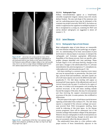
Figure 32.29 Lateral (a) and craniocaudal (b) views of the
humerus. It is imperative to obtain orthogonal radiographs of Regardless of the disease process, there are specific radio-
any suspected trauma case. Notice on the lateral view (a) that graphic changes identified with joint pathology. These
the fracture is more difficult to detect while on the craniocaudal include (Figure 32.39): soft tissue swelling, changes in the
view, there is an obvious large oblique middiaphyseal fracture joint space, changes in the subchondral bone (#), articular
with lateral displacement and overriding of the segments.
mineralization (Ω), joint incongruency (*), osteophytes (α),
enthesophytes(π), and intraarticular gas [7, 54].
Soft tissue swelling is a common response to join injury
and may be intraarticular or periarticular. The joint carti-
lage, synovial fluid and membrane, and joint capsule are
all of soft tissue opacity and cannot be differentiated radio-
graphically [7, 54]. Soft tissue swelling will appear as
increased soft tissue opacity of the affected joint and loss of
normal intraarticular fat pads. If confined to the region of
the joint, it is likely to be intracapsular and involving the
synovial structures. If the soft tissue swelling extends
beyond the margins of the joint, this may be extracapsular
and thus involving the support structures/nonsynovial
structures of the joint. In some patients, this is difficult to
differentiate.
The joint space is the region of soft tissue opacity between
opposing subchondral bone surfaces [54]. The soft tissue
opacity is composed of the articular cartilage and associ-
ated synovial fluid. With intracapsular swelling, there
could be widening of this joint space. As the disease
process progresses, there will be subsequent loss of the
articular cartilage with resultant collapse of the joint space.
However, because radiographs of small animal patients are
obtained non-weight bearing, changes in joint space size
may not accurately reflect the disease status [7].
Figure 32.30 Classification of the five most common types of
Salter–Harris fracture. (a) Normal, (b) Type I, (c) Type II, (d) Type The subchondral bone plate is a thin uniform radio-
III, (e) Type IV, (f) Type V. paque section of bone immediately adjacent to the