
Page 528 - Small Animal Clinical Nutrition 5th Edition
P. 528
546 Small Animal Clinical Nutrition
history are compatible with acute pancreatitis; however,
VetBooks.ir abdominal radiographs, ultrasound and laboratory evidence
supporting a diagnosis of pancreatitis are typically lacking.
The term pseudopancreatitis has been used to describe the
clinical manifestations associated with hypertriglyceridemia
(Ford, 1993, 1996).
Lipemia retinalis, a condition characterized by pale pink reti-
nal arterioles and venules, is an incidental finding seen on fun-
duscopic examination of lipemic dogs and cats (Figure 28-4).
This condition does not affect vision. Laboratory analysis of
affected animals will verify extreme hypertriglyceridemia, typi-
cally greater than 1,000 mg/dl.
Sustained hypertriglyceridemia is a principal risk factor
among people and dogs for developing acute pancreatitis
(Brown and Goldstein, 1987; Ford, 1993; Armstrong and Ford,
1989; DeBowes, 1987; Sanfey and Cameron, 1985; Whitney et
al, 1987; Williams, 1995). Dogs with acute abdominal pain and
vomiting should be evaluated for hyperchylomicronemia at the
time of presentation and during the recovery phase when food
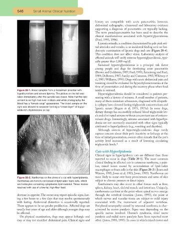
Figure 28-1. Blood samples from a Doberman pinscher with intake is restored.
hypothyroidism and severe lipemia. The picture on the left was Hypertriglyceridemia should be considered in patients pre-
taken immediately after the sample was drawn. Note that the lipid senting with a history of seizures. A small number of patients,
content is so high that even in black and white photography the many of them miniature schnauzers, diagnosed with idiopath-
blood has a “tomato soup” appearance. The blood sample on the
ic epilepsy have elevated fasting triglyceride concentrations and
right was allowed to separate forming a “cream layer” of triglyc-
lipemic serum (Rogers et al, 1975, 1975a). In some dogs,
eride-rich chylomicrons on top.
dietary therapy has successfully reduced blood triglyceride lev-
els and eliminated seizures without concomitant use of anticon-
vulsant drugs. Interestingly, seizures associated with hyperlipi-
demia are not necessarily associated with other signs typically
attributed to hyperlipidemia (e.g., vomiting and diarrhea).
Although owners of hypertriglyceridemic dogs rarely
express concern about their pet’s inactivity or lethargy at the
time of initial presentation, owners often remark that the pet’s
activity level increased as a result of lowering circulating
triglyceride levels. a
Cats with Hyperlipidemia
Clinical signs in hyperlipidemic cats are different than those
reported to occur in dogs (Table 28-1). The most common
clinical finding in affected cats is cutaneous xanthoma, a pain-
less, raised lesion caused by accumulation of lipid-laden
macrophages or foam cells in the skin (Figure 28-2) (Jones and
Watson, 1995; Jones et al, 1983; Jones, 1995). Xanthomas are
Figure 28-2. Xanthomas on the pinna of a cat with hyperlipidemia. most likely to occur over bony prominences and areas of skin
Xanthomas are tumors composed of lipid-laden foam cells, which subject to chronic pressure or direct injury.
are histiocytes containing cytoplasmic lipid material. These lesions Xanthomata may also occur in other tissues such as liver,
resolved with use of a low-fat, high-fiber food. spleen, kidney, heart, skeletal muscle and intestines. Uniquely,
xanthomata can form at the point where spinal nerves emerge
decrease in appetite.The owner may report episodic signs, last- through the vertebral foramina (Jones, 1993), the point at
ing a few hours to a few days that may resolve spontaneously which nerves and vascular tissue are subject to mild injury
with fasting. Abdominal distention is occasionally reported. associated with the movement of adjacent vertebrae.
There appears to be no gender predilection. Affected dogs are Peripheral neuropathy caused by neuronal xanthoma is char-
usually four years of age and older although younger dogs may acterized by motor paralysis. Signs vary depending on the
be affected. specific nerves involved. Horner’s syndrome, tibial nerve
On physical examination, dogs may appear lethargic and paralysis and radial nerve paralysis have been reported most
may or may not manifest abdominal pain. Clinical signs and often (Jones, 1993, 1995). In cases in which mixed motor and