
Page 543 - Small Animal Clinical Nutrition 5th Edition
P. 543
562 Small Animal Clinical Nutrition
transport glucose across the lipid bilayers into the cytosol. At
VetBooks.ir least five glucose transporters have been described, to date, in
people, each having a different affinity for glucose. GLUT 1
and GLUT 3 are present in all tissues and mediate basal glu-
cose uptake and neuronal uptake of glucose, respectively.
GLUT 2 is the major glucose transporter in beta and hepatic
cells. It has a low affinity for glucose, and acts as a transporter
during periods of hyperglycemia. GLUT 5 is found on the
brush border of human small intestinal cells and is mainly a
fructose transporter. GLUT 4 is found intracellularly in
insulin-dependent tissues, most notably skeletal muscle and
adipose tissue. Activation of the insulin signaling cascade
results in movement of GLUT 4 transporters to the cell surface
where the transporter facilitates glucose entry into cells (James
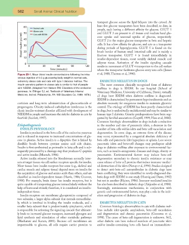
Figure 29-1. Mean blood insulin concentrations following the intra- et al, 1988; Thorens et al, 1990).
venous injection of 0.5 g glucose/kg body weight in normal cats,
extremely obese cats and cats with type I diabetes mellitus. The DIABETES MELLITUS IN DOGS
insulin secretory pattern in obese cats is similar to that of people The most common clinically recognized form of diabetes
with NIDDM. (Adapted from Nelson RW. Disorders of the endocrine mellitus in dogs is IDDM. In our hospital (School of
pancreas. In: Ettinger SJ, ed. Textbook of Veterinary Internal Veterinary Medicine, University of California, Davis), virtually
Medicine, 3rd ed. Philadelphia, PA: WB Saunders Co, 1989; 1676.)
all dogs have IDDM when diabetes mellitus is diagnosed.
IDDM is characterized by permanent hypoinsulinemia and an
corticism and long-term administration of glucocorticoids or absolute necessity for exogenous insulin to maintain glycemic
progestagens. Obesity-induced carbohydrate intolerance is the control. The etiology of IDDM has been poorly characterized
classic insulin-resistant disorder affiliated with development of in dogs, but is undoubtedly multifactorial and may be similar to
NIDDM in people and increases the risk for diabetes in cats by human type I diabetes. Genetic predispositions have been sug-
fourfold (Scarlett, 1997). gested by familial associations (Guptill, 1999; Hess et al, 2000).
Common histologic abnormalities in dogs include a reduction
Etiopathogenesis in the number and size of pancreatic islets, a decrease in the
INSULIN PHYSIOLOGY number of beta cells within islets and beta-cell vacuolation and
Insulin is produced in the beta cells of the endocrine pancreas degeneration. In some dogs, an extreme form of the disease
and is released in response to increased concentrations of glu- may occur, represented by a congenital absolute deficiency of
cose in plasma. Active insulin is a dipeptide that is linked by beta cells and pancreatic islet hypoplasia or aplasia. Less severe
disulfide bonds between cysteine amino acid side chains. pancreatic islets and beta-cell changes may predispose adult
Insulin is first synthesized as proinsulin in beta cells and is sub- dogs to diabetes mellitus after exposure to environmental fac-
sequently processed by a cleavage step that produces C-peptide tors, such as insulin-antagonistic diseases and drugs, obesity or
and active insulin (Muench, 1986). pancreatitis. Environmental factors may induce beta-cell
Active insulin released into the bloodstream normally inter- degeneration secondary to chronic insulin resistance or may
acts at target tissues via cell surface receptors specific for insulin. cause release of beta-cell proteins that induce immune-mediat-
Most tissues have insulin receptors but some (e.g., skeletal and ed destruction of the islets (Nerup et al,1994).Studies designed
cardiac muscle and adipose tissue) depend more on insulin for to detect anti-beta-cell autoantibodies in diabetic dogs have
the acquisition of glucose and amino acids than others, and are been conflicting; they were identified in newly-diagnosed dia-
classified as insulin-dependent tissues (Harris, 1986; Granner, betic dogs with IDDM in one study (Hoenig and Dawe, 1992)
1988). For example, brain tissue has insulin receptors, but is but not in another (Haines, 1986). Immune-mediated insulitis
quite capable of transporting glucose intracellularly without the has also been described in diabetic dogs (Alejandro et al, 1988).
help of hormonal stimuli; therefore, it is considered an insulin- Seemingly, autoimmune mechanisms, in conjunction with
independent tissue. genetic and environmental factors, may play a role in the initi-
Insulin receptors are membrane glycoproteins composed of ation and progression of diabetes in dogs.
two subunits; a larger alpha subunit that extends extracellular-
ly, which is involved in binding the insulin molecule, and a DIABETES MELLITUS IN CATS
smaller beta subunit that is predominately cytoplasmic, which Common histologic abnormalities in cats with diabetes mel-
is responsible for activating the signaling cascade that ultimate- litus include islet-specific amyloidosis, beta-cell vacuolation
ly leads to increased glucose transport, increased glycogen and and degeneration and chronic pancreatitis (Goossens et al,
lipid synthesis and stimulation of other metabolic pathways 1998). The cause of beta-cell degeneration is unknown. Still,
(Masharani and Karam, 2001). Because cell membranes are other diabetic cats have reduced numbers of pancreatic islets
impermeable to glucose, all cells require carrier proteins to and/or insulin-containing beta cells based on immunohisto-