
Page 573 - Small Animal Clinical Nutrition 5th Edition
P. 573
594 Small Animal Clinical Nutrition
et al, 1987) have also been associated with tumors cells.
VetBooks.ir Increased glycolysis in tumors may also be a consequence of
hexokinase redistribution in subcellular and mitochondrial
compartments, or the altered ability of mitochondria to metab-
olize substrates other than carbohydrates or their derivatives for
energy (Ristow, 2006). Numerous mechanisms have been pro-
posed and studied to clarify the altered mitochondrial metabo-
lism in tumor cells.
Regardless of the exact mechanism(s) involved, increasing
the rate of glycolysis in tumor cells promotes tumor cell growth
in several species including dogs (Ogilvie and Vail, 1992; Heber
et al, 1986), forming lactate as an end product. The host must
then expend energy to convert lactate to glucose via the Cori
cycle, resulting in a net energy gain by the tumor and a net
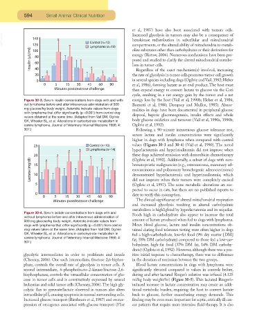
Figure 30-3. Serum insulin concentrations from dogs with and with- energy loss by the host (Vail et al, 1990b; Heber et al, 1986;
out lymphoma before and after intravenous administration of 500 Bozzetti et al, 1980; Dempsey and Mullen, 1985). Abnor-
mg glucose/kg body weight. Asterisks indicate values from dogs
malities in dogs have been documented in peripheral glucose
with lymphoma that differ significantly (p <0.001) from control dog
disposal, hepatic gluconeogenesis, insulin effects and whole
values obtained at the same time. (Adapted from Vail DM, Ogilvie
body glucose oxidation and turnover (Vail et al, 1990a, 1990b;
GK, Wheeler SL, et al. Alterations in carbohydrate metabolism in
canine lymphoma. Journal of Veterinary Internal Medicine 1990; 4: Ogilvie et al, 1992).
307.) Following a 90-minute intravenous glucose tolerance test,
serum lactate and insulin concentrations were significantly
higher in dogs with lymphoma when compared with control
values (Figures 30-3 and 30-4) (Vail et al, 1990). The noted
hyperlactatemia and hyperinsulinemia did not improve when
these dogs achieved remission with doxorubicin chemotherapy
(Ogilvie et al, 1992). Additionally, a subset of dogs with non-
hematopoietic malignancies (e.g., osteosarcoma, mammary ad-
enocarcinoma and pulmonary bronchogenic adenocarcinoma)
demonstrated hyperlactatemia and hyperinsulinemia, which
did not improve when their tumors were completely excised
(Ogilvie et al, 1997). The same metabolic alterations are sus-
pected to occur in cats, but there are no published reports to
date to verify this assumption.
The clinical significance of altered mitochondrial respiration
and increased glycolysis resulting in altered carbohydrate
metabolism is highlighted by hyperlactatemia and its sequelae.
Figure 30-4. Serum lactate concentrations from dogs with and Foods high in carbohydrate also appear to increase the total
without lymphoma before and after intravenous administration of
500 mg glucose/kg body weight. Asterisks indicate values from amount of lactate produced when fed to dogs with lymphoma.
dogs with lymphoma that differ significantly (p <0.001) from control Mean blood glucose, lactate and insulin concentrations ob-
dog values taken at the same time. (Adapted from Vail DM, Ogilvie tained during food tolerance testing were often higher in dogs
GK, Wheeler SL, et al. Alterations in carbohydrate metabolism in fed a high-carbohydrate, low-fat food (9% dry matter [DM]
canine lymphoma. Journal of Veterinary Internal Medicine 1990; 4: fat, 58% DM carbohydrate) compared to those fed a low-car-
307.)
bohydrate, high-fat food (37% DM fat, 14% DM carbohy-
drate) (Ogilvie et al, 1992). However, although there was a pos-
glycolytic intermediates in order to proliferate and invade itive initial response to chemotherapy, there was no difference
(Chesney, 2006). One such intermediate, fructose-2,6-biphos- in the duration of remission between the two groups.
phate, controls the overall rate of glycolysis in tumor cells. A Blood lactate concentrations in dogs with lymphoma were
second intermediate, 6-phosphofructo-2-kinase/fructose-2,6- significantly elevated compared to values in controls before,
bisphosphatase, controls the intracellular concentration of glu- during and after lactated Ringer’s solution was infused (4.125
cose in tumor cells and is constitutively expressed by several ml/kg body weight/hr) (Figure 30-5). This lactated Ringer’s-
leukemias and solid tumor cells (Chesney, 2006).The high gly- induced increase in lactate concentration may create an addi-
colytic flux to pyruvate/lactate observed in tumors also alters tional metabolic burden, requiring the host to convert lactate
intracellular pH, causing apoptosis in normal surrounding cells. back to glucose, further exacerbating energy demands. This
Increased glucose transport (Birnbaum et al, 1987) and overex- finding may be even more important for septic, critically ill can-
pression of oncogenes associated with glucose transport (Flier cer patients that require more intensive fluid therapy. It is also