
Page 591 - Small Animal Clinical Nutrition 5th Edition
P. 591
Adverse Reactions to Food 613
sitivity, which has recently shown some promise (Arslan et al, Table 31-1. Gastrointestinal barriers to ingested food antigens.
2006; Gaschen et al, 2008).
VetBooks.ir Risk Factors Physiologic barriers
Breakdown ingested antigens
Risk factors for adverse food reactions in animals are currently Gastric acid and pepsin
unknown but may include: 1) certain foods or food ingredients Pancreatic enzymes
Intestinal enzymes
(see below), 2) poorly digestible proteins, 3) any disease that Intestinal epithelial cell lysozyme activity
increases intestinal mucosal permeability (e.g.,viral enteritis),4) Block penetration of ingested antigens
selective IgA deficiency, 5) genetic predisposition, 6) age (six Unstirred water layer
Intestinal mucous coat (glycocalyx)
months to four years) and 7) concurrent allergic disease. Intestinal microvillous membrane composition
Intestinal peristalsis
Etiopathogenesis Immunologic barriers
Block penetration of ingested antigens
Normal Mucosal Barrier and Oral Tolerance Antigen-specific secretory IgA in gut lumen
Ingested food represents the greatest foreign antigenic load Clear antigens penetrating GI barrier
confronting the immune system.The defense against hypersen- Monocyte-macrophage system
Serum antigen-specific IgA and IgG
sitivity to food antigens includes an effective mucosal barrier
and oral tolerance generated by the cellular immune system of
GALT (Strombeck and Guilford,1991; Sampson,1993;Walk-
er, 1987; Murphy and Walker, 1991).
An important adaptation of the GI tract is the development
of a mucosal barrier that prevents the overwhelming uptake of
food antigens (Sampson, 1993; Walker, 1987; Murphy and
Walker, 1991). Efficient functioning of the mucosal barrier
excludes the majority of ingested antigens, thus minimizing
antigen exposure to GALT. The concept of a mucosal barrier
includes effective digestion, the mucous layer, intact and func-
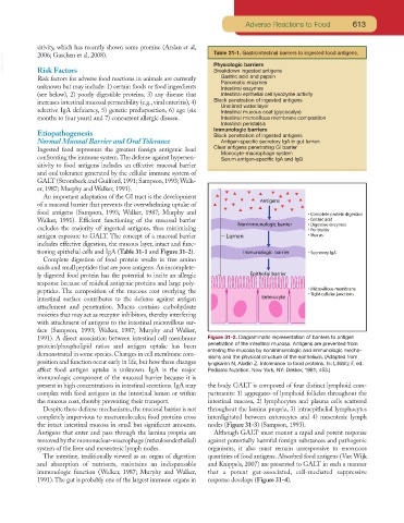
tioning epithelial cells and IgA (Table 31-1 and Figure 31-2).
Complete digestion of food protein results in free amino
acids and small peptides that are poor antigens. An incomplete-
ly digested food protein has the potential to incite an allergic
response because of residual antigenic proteins and large poly-
peptides. The composition of the mucous coat overlying the
intestinal surface contributes to the defense against antigen
attachment and penetration. Mucus contains carbohydrate
moieties that may act as receptor inhibitors, thereby interfering
with attachment of antigens to the intestinal microvillous sur-
face (Sampson, 1993; Walker, 1987; Murphy and Walker,
1991). A direct association between intestinal cell membrane Figure 31-2. Diagrammatic representation of barriers to antigen
protein/phospholipid ratios and antigen uptake has been penetration of the intestinal mucosa. Antigens are prevented from
demonstrated in some species. Changes in cell membrane com- entering the mucosa by nonimmunologic and immunologic mecha-
nisms and the physical structure of the epithelium. (Adapted from
position and function occur early in life, but how these changes Iyngkaren N, Abidin Z. Intolerance to food proteins. In: Lifshitz F, ed.
affect food antigen uptake is unknown. IgA is the major Pediatric Nutrition. New York, NY: Dekker, 1981; 453.)
immunologic component of the mucosal barrier because it is
present in high concentrations in intestinal secretions. IgA may the body. GALT is composed of four distinct lymphoid com-
complex with food antigens in the intestinal lumen or within partments: 1) aggregates of lymphoid follicles throughout the
the mucous coat, thereby preventing their transport. intestinal mucosa, 2) lymphocytes and plasma cells scattered
Despite these defense mechanisms, the mucosal barrier is not throughout the lamina propria, 3) intraepithelial lymphocytes
completely impervious to macromolecules; food proteins cross interdigitated between enterocytes and 4) mesenteric lymph
the intact intestinal mucosa in small but significant amounts. nodes (Figure 31-3) (Sampson, 1993).
Antigens that enter and pass through the lamina propria are Although GALT must mount a rapid and potent response
removed by the mononuclear-macrophage (reticuloendothelial) against potentially harmful foreign substances and pathogenic
system of the liver and mesenteric lymph nodes. organisms, it also must remain unresponsive to enormous
The intestine, traditionally viewed as an organ of digestion quantities of food antigens. Absorbed food antigens (Van Wijk
and absorption of nutrients, maintains an indispensable and Knippels, 2007) are presented to GALT in such a manner
immunologic function (Walker, 1987; Murphy and Walker, that a potent gut-associated, cell-mediated suppressive
1991).The gut is probably one of the largest immune organs in response develops (Figure 31-4).