
Page 609 - Small Animal Clinical Nutrition 5th Edition
P. 609
Adverse Reactions to Food 631
Dermatologic examination revealed marked traumatic and
complete alopecia with hyperpigmentation and erythema involv-
VetBooks.ir ing the periocular areas, inner pinnae, axillae, feet and ventral
abdomen (Figures 1 to 3). Small numbers of papules were found
on the ventral abdomen. Excoriations were present in the axillae
and periocular areas.
Assess the Food and Feeding Method
The dog was fed a variety of commercial dry foods; the client
changed brands frequently. The dry food was fed free choice.
Other food sources included occasional table food, commercial
canine biscuit treats, rawhide chews and flavored heartworm pre-
ventive medication, which was given monthly for nine months of
the year.
Questions
1. What are the primary diseases in the differential diagnosis of
this patient? What secondary diseases may be present?
2. What food and feeding method is appropriate for this patient?
3. How might the dog’s otitis externa correlate with the other
evidence of dermatologic disease?
Answers and Discussion
1. The primary diseases in the differential diagnosis include:
Atopic dermatitis. Most patients with atopic dermatitis have
pruritus and clinical disease at six months to three years of age.
A seasonal history also suggests atopic dermatitis. This dog’s
dermatologic problems began at five years of age and the pru-
ritus is nonseasonal, which is still compatible with atopic der-
matitis. Atopic dermatitis is more common than food allergy
but less common than flea allergy.
Adverse reaction to food (food allergy or food intolerance). The
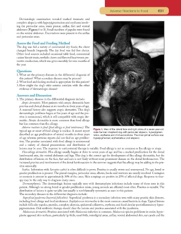
typical age at onset of food allergy is unclear. A recent report Figure 1. View of the lateral face and right pinna of a seven-year-old
male German shepherd dog with periocular alopecia, hyperpigmen-
described an age predilection of several months to three years tation, erythema and mild excoriations. The inner pinnal surface was
of age whereas previous reports did not find an age predilec- hyperpigmented, erythematous and alopecic.
tion. The pruritus associated with food allergy is nonseasonal
and a variety of clinical presentations and distribution of
lesions may be seen. The response to corticosteroid therapy is variable. Food allergy is not as common as flea allergy or atopy.
Flea-allergy dermatitis. Flea allergy usually begins at three to seven years of age and has a marked predilection for the dorsal
lumbosacral area, the ventral abdomen and legs. This dog is the correct age for development of flea-allergy dermatitis, but the
distribution of lesions on the face, feet and ears is not likely without more prominent disease on the dorsal lumbosacrum. The
increased pruritus and involvement of the dorsal lumbosacrum in the summer suggests that flea allergy may be adding to the pru-
ritus seasonally.
Scabies. Infestation with Sarcoptes scabiei is often difficult to prove. Pruritus is usually severe and nonseasonal. No age, breed or
gender predilection is present. The pinnal margins, periocular areas, elbows, hocks and ventrum are usually involved. Contagion
or zoonosis is present in approximately 30% of the cases. Skin scrapings are positive in 25% of affected dogs. Response to ther-
apy may be the only way to diagnose many cases.
Dermatophytosis. The dermatologic lesions typically seen with dermatophyte infections include many of those seen in this
patient. Although no strong breed or gender predilection exists, young animals are affected most often. Pruritus is variable. The
distribution of lesions is quite variable but usually is not bilaterally symmetric as seen in this patient.
The secondary diseases in the differential diagnosis include:
Superficial pyoderma (bacterial folliculitis). Superficial pyoderma is a secondary infection seen with many pruritic skin diseases,
including food allergy and food intolerance. Staphylococcus intermedius is the most common causal bacteria in dogs.Typical lesions
include follicular papules, pustules, complete alopecia, epidermal collarettes, erythema and focal circular postinflammatory hyper-
pigmentation. Oral antibiotic therapy should clear the lesions and pruritus associated with the pyoderma.
Malassezia dermatitis. Pruritus associated with Malassezia infection is common. Malassezia species proliferate in moist, hyper-
plastic apposed skin surfaces, particularly lip folds, nasal folds, interdigital areas, axillae, ventral abdominal skin, ear canals and the