
Page 631 - Small Animal Clinical Nutrition 5th Edition
P. 631
Skin and Hair Disorders 653
in the dermis, and assess the ability of IgE to fix allergen and
Table 32-6. Key nutritional factors for foods and supplements for
cause mast cell degranulation and subsequent vasodilatation. In
VetBooks.ir a well-controlled study using allergen mixes, 59% of dogs dogs and cats with inflammatory dermatoses.
Factors
Nutritional recommendations
responded to hyposensitization that was formulated on the
basis of intradermal testing results (Willemse et al, 1984). Omega-3 fatty acids (ALA, Supplements or foods should
EPA and/or DHA) initially provide 50 to 300 mg total
Intradermal allergy testing has several disadvantages (Hillier omega-3 fatty acids/kg body
and DeBoer, 2001). Negative intradermal results occur in some weight/day
Foods should contain between
dogs strongly suspected to have atopic dermatitis. Anti-
0.35 to 1.8% dry matter
inflammatory and antihistamine drugs must be withdrawn
before testing to prevent false-negative results. The test cannot Key: ALA = α-linolenic acid, EPA = eicosapentaenoic acid, DHA
= docosahexaenoic acid.
be performed on dogs with generalized dermatitis. Shaving of
the coat and sedation are usually required. Intradermal allergy
testing is time-consuming and not cost-effective when per-
formed infrequently.The usefulness of intradermal allergy test-
ing is also limited by lack of standardized allergy extracts and
no homogeneous criteria for the interpretation of results. Most
intradermal testing is performed at dermatologic referral cen-
ters because of these disadvantages. Intradermal testing for
food hypersensitivity is unreliable in animals with dermatolog-
ic disease (Chapter 31).
In vitro “allergy” tests measure serum concentrations of aller-
gen-specific IgE and avoid many of the disadvantages of intra-
dermal allergy tests (Codner and Griffin, 1996). In vitro tests
require only a serum sample; so they are readily available to pri-
vate practitioners and can be used on patients with generalized
dermatitis. Laboratories use several different techniques to
detect circulating IgE levels, including a radioallergosorbent
test (RAST), enzyme-linked immunosorbent assay (ELISA) or
liquid-phase enzyme immunoassay (EIA). Problems with in
vitro testing include poor reproducibility and a high false-pos-
itive rate (Codner and Lessard, 1993). Results to date suggest
that more than 60% of atopic dogs respond to hyposensitiza-
tion formulated on the basis of in vitro results (Anderson, 1993;
Sousa and Norton, 1990). In vitro testing is also available for
confirming flea-allergic dermatitis (Cook et al, 1996) but is
unreliable for diagnosing food hypersensitivity (Chapter 31).
Controversy continues over whether intradermal or in vitro
testing is the better method for confirming a diagnosis of atopic
dermatitis and for selecting allergens for hyposensitization
(DeBoer and Hillier, 2001). Furthermore, long-term studies are
needed to evaluate responses of allergic animals to hyposensiti-
zation based on both types of testing.
Risk Factors
Atopy (atopic state) is a genetically-predisposed tendency to
develop IgE-mediated allergy to environmental allergens (Oli-
vry et al, 2001c). Atopic disease is any clinical manifestation of
atopy including most commonly atopic dermatitis, atopic con-
junctivitis and/or atopic rhinitis (Olivry et al, 2001c). Atopic
dermatitis is a genetically predisposed inflammatory and pru-
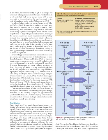
ritic allergic skin disease with characteristic clinical features Figure 32-3. Metabolic transformation of two major unsaturated
fatty acid families by desaturation and elongation.
(Olivry et al, 2001c). It is associated most commonly with IgE
antibodies to environmental allergens. Although the exact
mode of inheritance is unknown, strong breed predilection and clude Cairn terriers,West Highland white terriers, Scottish ter-
familial involvement in dogs indicate a genetically determined riers, wire-haired fox terriers, Boston terriers, Sealyham terriers,
cause. Canine breeds reported to be predisposed to atopy in- Lhasa apsos, Dalmatians, pugs, Irish setters, English setters,