Page 902 - Small Animal Clinical Nutrition 5th Edition
P. 902
934        Small Animal Clinical Nutrition


                                                                      urine culture and diagnostic imaging) are candidates.The lower
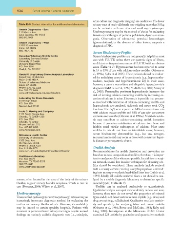
                   Table 46-9. Contact information for urolith analysis laboratories.
        VetBooks.ir  Antech Diagnostics – East                        urinary tract of nearly all female cats weighing more that 3.0 kg
                                                                      can be evaluated with one of several small rigid cystoscopes.
                   111 Marcus Ave.
                   Lake Success, NY 11042                             Urethrocystoscopy may be the method of choice for evaluating
                                                                      female cats with signs of periuria, pollakiuria, dysuria or stran-
                   800.872.1001
                                                                      guria. Observation of submucosal petechial hemorrhages
                   Antech Diagnostics – West                          (glomerulations), in the absence of other lesions, supports a
                   17672 Cowan Ave.                                   diagnosis of FIC.
                   Irvine, CA 92614
                   800.745.4725
                                                                      Serum Biochemistry Profiles
                   Canadian Veterinary Urolith Centre                 Serum biochemistry profiles are not generally helpful in most
                   Laboratory Services Division
                   University of Guelph                               cats with FLUTD unless there are systemic signs of illness,
                   95 Stone Road West                                 urolithiasis or frequent recurrences of FLUTD with no obvious
                   P.O. Box 3650                                      cause (Table 46-7). Hypercalcemia has been reported to occur
                   Guelph, ON N1H8J7
                                                                      in 14 to 35% of cats with calcium oxalate uroliths (Osborne et
                   Gerald V. Ling Urinary Stone Analysis Laboratory   al, 1996a; Kyles et al, 2005). These patients should be evaluat-
                   Department of Medicine                             ed for underlying causes of hypercalcemia (e.g., hyperparathy-
                   Room 3106 MSI-A
                   School of Veterinary Medicine                      roidism, neoplasia and hypervitaminosis D); in most cases,
                   Davis, CA 95616                                    however, a cause is not evident and idiopathic hypercalcemia is
                   Phone: 530.752.3228                                diagnosed (McClain et al, 1999; Midkiff et al, 2000; Savary et
                   Fax: 530.752.0414
                   www.vetmed.ucdavis.edu/vme/labs.htm                al, 2000). Presumably, persistent hypercalcemia increases the
                                                                      risk of forming calcium-containing uroliths by increasing ex-
                   Laboratory for Stone Research                      cretion of calcium in urine. However, it is possible that process-
                   81 Wyman Street
                   P.O. Box 129                                       es involved with formation of calcium-containing uroliths and
                   Newton, MA 02168                                   hypercalcemia are unrelated. Acidemia and serum total CO 2
                                                                      less than 18 mEq/l, were reported in 64% of non-azotemic cats
                   Louis C. Herring and Company
                   1111 South Orange Ave.                             with calcium oxalate uroliths and 92% of cats with concurrent
                   Orlando, FL 32806-1236                             azotemia and uroliths (Osborne et al, 1996a). Metabolic acido-
                   P.O. Box 2191                                      sis may contribute to calcium-containing urolith formation
                   Orlando, FL 32802
                   Phone: 407.841.770                                 because it promotes mobilization of calcium from bone and
                   Fax: 407.422.8896                                  inhibits renal tubular reabsorption of calcium. Most urate
                   www.herringlab.com                                 uroliths in cats do not have an identifiable cause; however,
                                                                      serum biochemistry abnormalities (e.g., low urea nitrogen,
                   Minnesota Urolith Center
                   University of Minnesota                            increased ammonia) may occur in those with concurrent hepat-
                   1352 Boyd Ave.                                     ic disease or portosystemic shunts.
                   St. Paul, MN 55108
                   Phone: 612.625.4221
                   Fax: 612.624.0751                                  Urolith Analysis
                   www.cvm.umn.edu/depts/minnesotaurolithcenter       Recommendations for urolith dissolution and prevention are
                                                                      based on mineral composition of uroliths; therefore, it is impor-
                   Urolithiasis Laboratory
                   P.O. Box 25375                                     tant to analyze uroliths whenever possible. In addition to surgi-
                   Houston, TX 77265-5375                             cal removal, several less invasive techniques for obtaining uro-
                   800.235.4846
                   www.urolithiasis-lab.com                           liths should be considered. These methods include retrieval
                                                                      with a urinary catheter, voiding urohydropropulsion and void-
                                                                      ing into an empty or plastic bead-filled litter box (Lulich et al,
                                                                      1993). Ideally, all uroliths retrieved from a cat should be ana-
                  masses, often located in the apex of the body of the urinary  lyzed by a urolith diagnostic laboratory to determine specific
                  bladder, suggest urinary bladder neoplasia, which is rare in  mineral type(s) (Table 46-9).
                  cats (Forrester, 2006; Wilson et al, 2007).           Uroliths can be analyzed qualitatively or quantitatively.
                                                                      Qualitative analysis uses spot tests to identify radicals and ions;
                  Urethrocystoscopy                                   however, these tests do not reveal the proportion of mineral
                  Transurethral cystoscopy (or urethrocystoscopy) is becoming an  types and do not detect certain mineral crystals (e.g., silica) and
                  increasingly important diagnostic method for evaluating the  drug crystals (e.g., sulfadiazine). Qualitative tests lack sensitiv-
                  urethra and urinary bladder of cats. However, its availability  ity and specificity for analyzing feline and canine uroliths
                  may be limited to certain specialty hospitals. Patients with  (Osborne et al, 1996; Bovee and McGuire, 1984; Ruby and
                  recurrent or persistent lower urinary tract signs despite normal  Ling, 1986). Investigators at the Minnesota Urolith Center
                  findings on routinely available diagnostic tests (i.e., urinalysis,  examined 223 uroliths by qualitative and quantitative methods

