
Page 37 - REVISED GP Fall 2021 - ready for posting
P. 37
of the antrum. Being prepared with a knowledge of anatomy allows Conflict of interests
the operator to anticipate how to leverage the instrument to follow the The author claims to have no financial interest, directly or indirectly, in any
contours of the antrum. A septa was encountered in the vicinity of the MB
root of #3. This was anticipated and would become the posterior wall of entity that is commercially related to the products mentioned in this paper.
the osseous graft. During the dissection, the membrane was released to The author reports that no conflict of interest exists.
the top of the septa and along the medial wall to an area superior to the
root of #4. This created a box bounded by the septa on the posterior, the References
medial wall on the medial side, and the root complex of #4 on the anterior. 1. Tatum OH: Lecture presented at Alabama Implant Study Group, Birmingham
Alabama. 1977.
The sinus membrane was elevated using a resorbable collagen 2. Misch CE. (2008) Contemporary Implant Dentistry. (3 Ed.) Mosby Elsevier.
rd
membrane, Biomend Extend (Zimmer/BIOMET, Warsaw, Ind). This p. 929.
allowed the graft space to be inspected with a directed light source 3. Boyne PJ, James RI Grafting of the maxillary sinus floor with autogenous
and the extent of the lifted membrane was verified. The floor of the marrow and bone. J.Oral Surg 38:613-616. 1980.
internal osseous bed was scratched with an explorer to stimulate bone 4. Riben C., Thor A. The Maxillary Sinus Membrane Elevation Procedure:
production activity. 14 Augmentation of Bone around Dental Implants without Grafts—A Review of a
Surgical Technique. Int J Dent, 2012; 2012: 105483.
A mixture of RegenerOss Allograft Putty Plus mineralized bone 5. Kalvyas D., et. al., Int J Implant Dent. Dec. 4:32.2018.
(Zimmer/BIOMET, Warsaw, Ind) was serially delivered to the 6. Lin YH. et. al., The influence of the sinus membrane thickness upon
osteotomy and introduced into the prepared bed. Aliquots of bone membrane perforation during lateral window sinus augmentation. Clin Oral
graft were brought to the osteotomy on the large flat end of a periosteal Implants Res. 2016 May: 27(5): 612-7.
elevator and delivered into the antrum with a plastic instrument. As the 7. Testori T. Maxillary sinus surgery. Anatomy and advanced diagnostic
imaging. J Implant and Reconstructive Dent. 2011; 2: 6-14.
area began to fill, the graft was distributed toward the media wall, and rd
then to the distal and mesial extensions of the intended graft site. A 8. Misch CE. (2008) Contemporary Implant Dentistry. (3 Ed.) Mosby Elsevier.
p. 951.
total of 0.5 cc’s of bone graft was introduced.
9. Misch CE. (2008) Contemporary Implant Dentistry. (3 Ed.) Mosby Elsevier.
rd
p. 949 .
After gentle packing to remove air inclusions, another collagen 10. Misch CE. et. al., Indications for and classifications of sinus bone grafts.
membrane was placed over the osteotomy and pressed for closure. The In Jensen OT, editor: The sinus bone graft. ed 2, Chicago, 2006, Quintessence.
area was irrigated and the flap was returned to the osseous bed. The flap 11. Jeong TM., Lee JK. The Efficacy of the Graft materials after sinus elevation:
was closed with Teflon 4-0 suture without tension. Retrospective Comparative Study Using Panoramic Radiography. Maxillofac
Plast Reconstr Surg 2014 Jul; 36(4):146-153.
The patient was given postoperative instructions which included 12. Triplett RG, et. al., Pivotal, randomized, parallel evaluation of recombinant
warnings about blowing his nose and sneezing. He was instructed to human bone morphogenetic protein-2/absorbable collagen sponge and
continue with two pre-operative prescriptions, Amoxicillin 500 mg 1 autogenous bone graft for maxillary sinus floor augmentation. J Oral Maxillofac
q 6 h, 28 tablets and a medrol dose pack (Methylprednisolone - 4 mg Surg. 2009 Sep; 67(9):1947-60.
tablets) (Sandoz, Princeton, NJ). Both prescriptions were started the day 13. Hallman M, Thor A. Bone substitutes and growth factors as an alternative/
before the procedure and continued for several days after the procedure. complement to autogenous bone for grafting in implant dentistry. Periodontol
2000. 2008; 47():172-92.
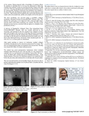
The case was followed for several months (Figure 10), however, due to 14. Misch CE. (2008) Contemporary Implant Dentistry. (3 Ed.) Mosby
rd
patient scheduling the implant was not placed until 12 months after the Elsevier p. 946.
sinus lift (Figures 11-13)
Dr. DiDonato is in
private practice in
Rochester, NY. He is
a past president of the
New York State Academy
of General Dentistry and
is currently serving as its
treasurer.
10 Figure 10. Sinus graft at 6 months with 11 Figure 11. Sinus graft at 1 year, implant
Sinus graft at 1 year, implant placement
faint indication of original sinus floor (dark
Sinus graft at 6 months with faint indication of original sinus floor (dark placement.
arrow) and new sinus floor (white arrow)
arrow) and new sinus floor (white arrow). Final restoration occlusal view 13
12 Figure 12. Final restoration. Figure 13. Final restoration occlusal view. www.nysagd.org l Fall 2021 l GP 37
Final restoration