
Page 4 - 48_Freemartinism
P. 4
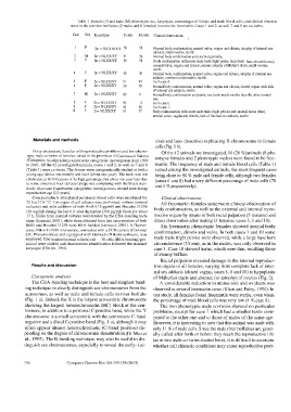
Table 1. Female (F) and male (M) phenotypic sex, karyotype, percentages of female and male blood cells, and clinical observa-
                              tions in the ten river buffaloes (2 males and 8 females) found to be freemartin. Cases 1 and 2, as well 7 and 8 are co-twins.
                               Case  Sex  Karyotype  F cells  M cells  Clinical observations
                                                                              (%)
                                1   F               50    50    Normal body conformation, normal vulva, vagina and clitoris, atrophy of internal sex
                                        2n = 5O,XX/XY
                                                                adducts, small ovaries, sterile.
                               2    M   2n = 5O,XX/XY  41  59   Normal body conformation and external genitalia.
                               3    F   2n = 5O,XX/XY  50  50   Body conformation with some male traits (tight pelvis, large horn base circumference),
                                                                normal vulva, vagina and clitoris, serious atrophy of Muller's ducts, small ovaries,
                                                                sterile.
                               4    F   2n = 5O,XX/XY  50  50   Normal body conformation, normal vulva, vagina and clitoris, atrophy of internal sex
                                                                adducts, ovaries not detectable, sterile.
                               5    F   2n = 5O,XX/XY  51  49   As in case 4.
                               6    F   2n = 5O,XX/XY  30  70   Normal body conformation, normal vulva, vagina and clitoris, closed vagina with lack
                                                                of internal sex adducts, sterile.
                               7    M   2n = 5O,XX/XY  89  11   Normal body conformation and penis, one testis much smaller than the other normal
                                                                one.
                               8    F  2n = 5O,XX/XY  96  4     As in case 6.
                               9    F  2n = 5O,XX/XY  50  50    As in case 1.
                               10   F  2n= 5O,XX/XY  63   37    Body conformation with some male traits (tight pelvis) and unusual horns (thin),
                                                                normal vulva, vagina and clitoris, lack of internal sex adducts, sterile.
               Materials and methods                             tive) and late- (inactive) replicating X chromosome in female
                                                                 cells (Fig. 1 b).
               Forty phenotypic females with reproductive problems and two pheno-
                                                                   Of the 42 animals we investigated, 10 (24 %)animals (8 phe-
            typic male co-twins of females, raised in the provinces of Caserta and Salerno
                                                                notypic females and 2 phenotypic males) were found to be free-
            (Campania, Southern Italy) underwent cytogenetic investigation from 1998
            to 2003. Of the 42 investigated animals, cases 1 and 2, as well as 7 and 8  martin. The frequency of male and female blood cells (Table 1)
            (Table 1) were co-twins. The former were cytogenetically studied at both a  varied among the investigated animals, the most frequent cases
            young age (about one month) and later (about one year). The male was not  being close to 50 % male and female cells, although two females
            eliminated at birth because of its high genealogy, but about one year later due  (cases 6 and 8) had a very different percentage of male cells (70
            to some observed hoof defects (shape not complying with the breed stan-
                                                                and 4 %,respectively).
            dard). Also case 8 underwent cytogenetic investigation a second time during
            reproductive age (2.5 years).
              Concanavalin A-stimulated peripheral blood cells were incubated for  Clinical observations
            72 h at 37.8 ° C. Two types of cell cultures were performed: without (normal  All freemartin females underwent clinical observation of
            cultures) and with addition of both BrdU (15 gg/ml) and Hoechst 33258
                                                                body conformation, as well as the external and internal repro-
            (30 wg/ml) during the last 6 b after thymidine (300 pg/ml) block for about
            17 h. Slides from normal cultures were treated by the CBA-banding tech-  ductive organs by means of both rectal palpation (5 females) and
            nique (Iannuzzi, 2003), while those obtained from late incorporation of both  direct observation after mating (3 females; cases 3, 6 and 10).
            BrdU and Hoechst 33258 were RBA-banded (Iannuzzi, 2003). A fluores-  Six freemartin phenotypic females showed normal body
            cence Nikon E-1000 microscope, connected with a CCD camera (Coolsnap
            CF, Photometrics) and equipped with Genicon (Nikon) software, was  conformation, clitoris and vulva. In both cases 3 and 10 some
            employed. One  hundred (normal cultures) and  30 cells (RBA-banding) per  male traits (tight pelvis) were observed, while a large base horn
            animal were studied and chromosome identification followed the standard  circumference (35 em), as in the males, was only observed in
            karyotype (CSKBB, 1994).                            case 3. Case 10 showed horns, which were thin, recalling those
                                                                of swamp buffalo.
                                                                   Rectal palpation revealed damage in the internal reproduc-
              Results and discussion                            tive organs of all females, varying from complete lack of inter-
                                                                nal sex adducts (closed vagina, cases 6, 8 and 10) to hypoplasia
              Cytogenetic analysis                              of Múllerian ducts and absence (or atrophy) of ovaries (Fig. 2).
              The CBA-banding technique is the best and simplest band-  A considerable reduction in uterus size and oviducts was
           ing technique to clearly distinguish sex chromosomes from the  observed in several freemartin cows (Khan and Foley, 1994). In
           autosomes, as well as male and female cells in river buffalo  our study, all females found freemartin were sterile, even when
           (Fig. 1 a). Indeed, the X is the largest acrocentric chromosome  the percentage of male blood cells was very low (4 %,case 8).
           showing the largest heterochromatin (HC) block at the cen-  The two phenotypic male co-twins showed no particular
           tromere, in addition to a proximal C-positive band, while the Y  problems, except for case 7 which had a smaller testis com-
           chromosome is a small acrocentric with the centromere C-band  pared to the other one and to those of males of the same age.
           negative and a distal C-positive band (Fig. 1 a), although it may  However, it is interesting to note that this animal was male with
           often appear almost heterochromatic (C-band positive) de-  only 11 % of male cells. Since the male river buffaloes are gener-
           pending on the degree of chromosome denaturation (Di Meo et  ally culled after birth or before they reach the reproductive life
           al., 1995). The R-banding technique may also be useful to dis-  (as in two male co-twins studied here), it is difficult to ascertain
           tinguish sex chromosomes, especially to reveal the early- (ac-  whether cell chimeric conditions may cause reproductive prob-
           356              Cytogenet Genome Res 108:355-358 (2005)