
Page 232 - Basic _ Clinical Pharmacology ( PDFDrive )
P. 232
218 SECTION III Cardiovascular-Renal Drugs
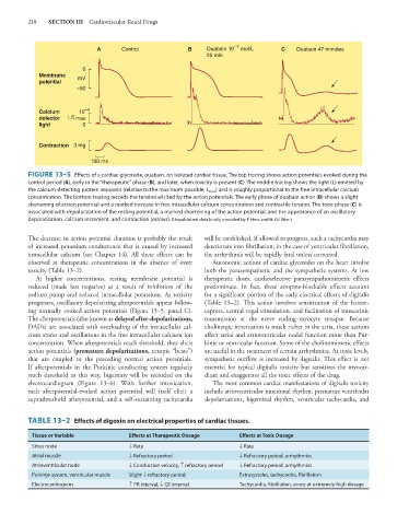
A Control B Ouabain 10 –7 mol/L C Ouabain 47 minutes
25 min
0
Membrane mV
potential
–50
Calcium 10 –4
detector L/L max
light 0
Contraction 3 mg
100 ms
FIGURE 13–5 Effects of a cardiac glycoside, ouabain, on isolated cardiac tissue. The top tracing shows action potentials evoked during the
control period (A), early in the “therapeutic” phase (B), and later, when toxicity is present (C). The middle tracing shows the light (L) emitted by
the calcium-detecting protein aequorin (relative to the maximum possible, L max ) and is roughly proportional to the free intracellular calcium
concentration. The bottom tracing records the tension elicited by the action potentials. The early phase of ouabain action (B) shows a slight
shortening of action potential and a marked increase in free intracellular calcium concentration and contractile tension. The toxic phase (C) is
associated with depolarization of the resting potential, a marked shortening of the action potential, and the appearance of an oscillatory
depolarization, calcium increment, and contraction (arrows). (Unpublished data kindly provided by P. Hess and H. Gil Wier.)
The decrease in action potential duration is probably the result will be established. If allowed to progress, such a tachycardia may
of increased potassium conductance that is caused by increased deteriorate into fibrillation; in the case of ventricular fibrillation,
intracellular calcium (see Chapter 14). All these effects can be the arrhythmia will be rapidly fatal unless corrected.
observed at therapeutic concentrations in the absence of overt Autonomic actions of cardiac glycosides on the heart involve
toxicity (Table 13–2). both the parasympathetic and the sympathetic systems. At low
At higher concentrations, resting membrane potential is therapeutic doses, cardioselective parasympathomimetic effects
reduced (made less negative) as a result of inhibition of the predominate. In fact, these atropine-blockable effects account
sodium pump and reduced intracellular potassium. As toxicity for a significant portion of the early electrical effects of digitalis
progresses, oscillatory depolarizing afterpotentials appear follow- (Table 13–2). This action involves sensitization of the barore-
ing normally evoked action potentials (Figure 13–5, panel C). ceptors, central vagal stimulation, and facilitation of muscarinic
The afterpotentials (also known as delayed after-depolarizations, transmission at the nerve ending–myocyte synapse. Because
DADs) are associated with overloading of the intracellular cal- cholinergic innervation is much richer in the atria, these actions
cium stores and oscillations in the free intracellular calcium ion affect atrial and atrioventricular nodal function more than Pur-
concentration. When afterpotentials reach threshold, they elicit kinje or ventricular function. Some of the cholinomimetic effects
action potentials (premature depolarizations, ectopic “beats”) are useful in the treatment of certain arrhythmias. At toxic levels,
that are coupled to the preceding normal action potentials. sympathetic outflow is increased by digitalis. This effect is not
If afterpotentials in the Purkinje conducting system regularly essential for typical digitalis toxicity but sensitizes the myocar-
reach threshold in this way, bigeminy will be recorded on the dium and exaggerates all the toxic effects of the drug.
electrocardiogram (Figure 13–6). With further intoxication, The most common cardiac manifestations of digitalis toxicity
each afterpotential-evoked action potential will itself elicit a include atrioventricular junctional rhythm, premature ventricular
suprathreshold afterpotential, and a self-sustaining tachycardia depolarizations, bigeminal rhythm, ventricular tachycardia, and
TABLE 13–2 Effects of digoxin on electrical properties of cardiac tissues.
Tissue or Variable Effects at Therapeutic Dosage Effects at Toxic Dosage
Sinus node ↓ Rate ↓ Rate
Atrial muscle ↓ Refractory period ↓ Refractory period, arrhythmias
Atrioventricular node ↓ Conduction velocity, ↑ refractory period ↓ Refractory period, arrhythmias
Purkinje system, ventricular muscle Slight ↓ refractory period Extrasystoles, tachycardia, fibrillation
Electrocardiogram ↑ PR interval, ↓ QT interval Tachycardia, fibrillation, arrest at extremely high dosage