
Page 162 - Atlas of Histology with Functional Correlations
P. 162
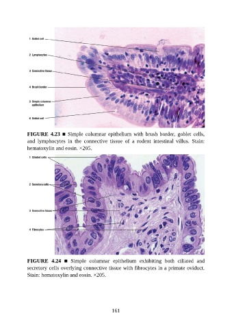
FIGURE 4.23 ■ Simple columnar epithelium with brush border, goblet cells,
               and  lymphocytes  in  the  connective  tissue  of  a  rodent  intestinal  villus.  Stain:

               hematoxylin and eosin. ×205.


               FIGURE  4.24  ■  Simple  columnar  epithelium  exhibiting  both  ciliated  and
               secretory cells overlying connective tissue with fibrocytes in a primate oviduct.
               Stain: hematoxylin and eosin. ×205.


                                                          161